Right auricular and right ventricular mass with tamponade, likely hemangiosarcoma, in a 13 year old FS Cairn Terrier

Case Study

Right auricular and right ventricular mass with tamponade, likely hemangiosarcoma, in a 13 year old FS Cairn Terrier

A 13-year-year FS Cairn terrier with a history PuPd was presented for evaluation of an episode of collapse. Pale mucous membranes were present on physical examination and serum biochemistry showed elevated ALP activity (1853).

A 13-year-year FS Cairn terrier with a history PuPd was presented for evaluation of an episode of collapse. Pale mucous membranes were present on physical examination and serum biochemistry showed elevated ALP activity (1853).

DX

right auricular and right ventricular mass. Stable gallbladder mucocele

Image Interpretation

Exam of the cranial abdomen demonstrated excessive liver size, swollen contour, with conserved uniform architecture. Parenchymal echogenicity was diffusely isoechoic to the spleen and falciform fat. The gallbladder in this patient revealed striating bile, overdistention consistent with gallbladder mucocele.  No peripheral inflammation was noted.  These nonspecific changes may be idiopathic or breed specific, or associated with hormonal imbalance (Diabetes, Hypothyroidism, Cushing’s disease.) 

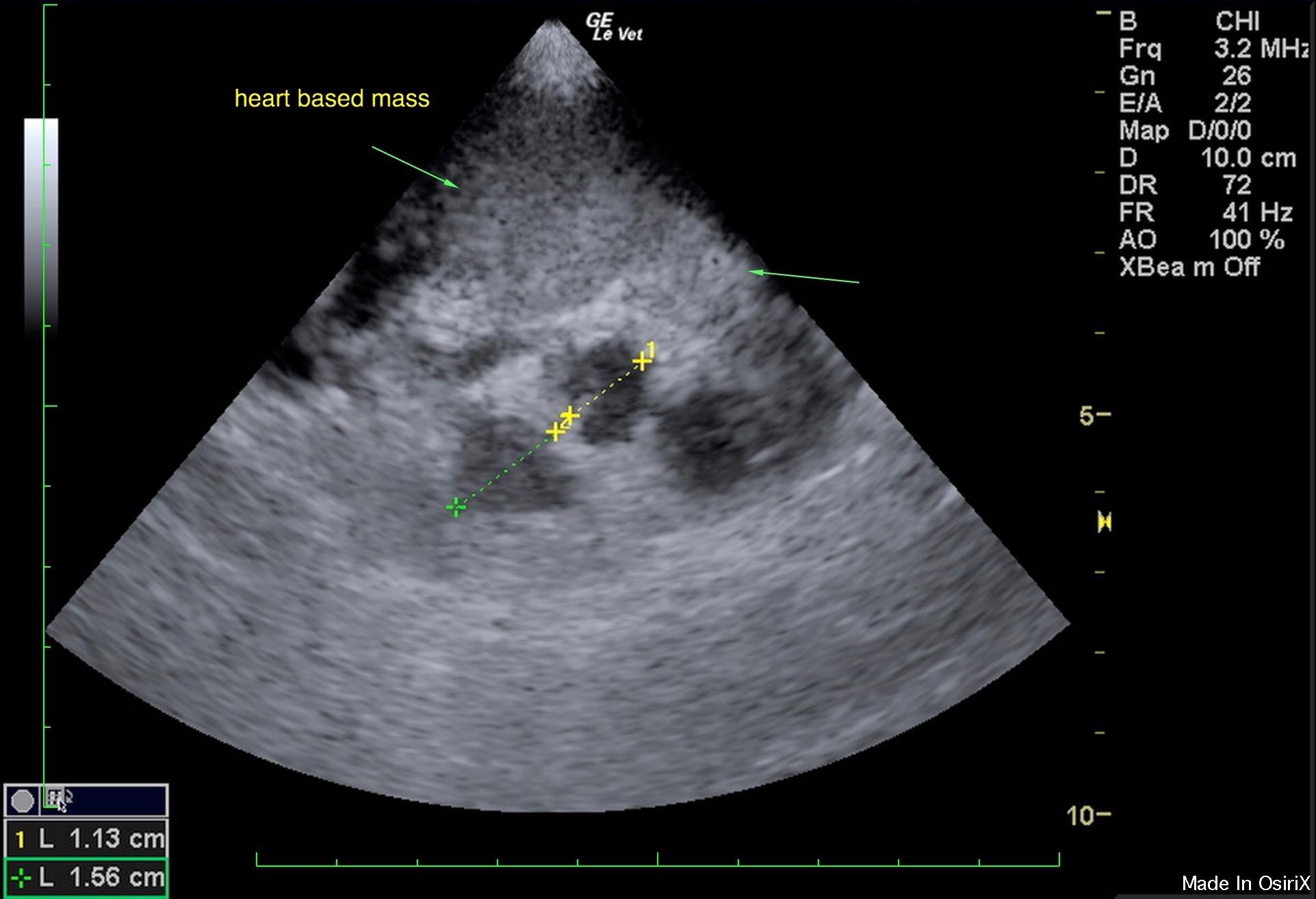
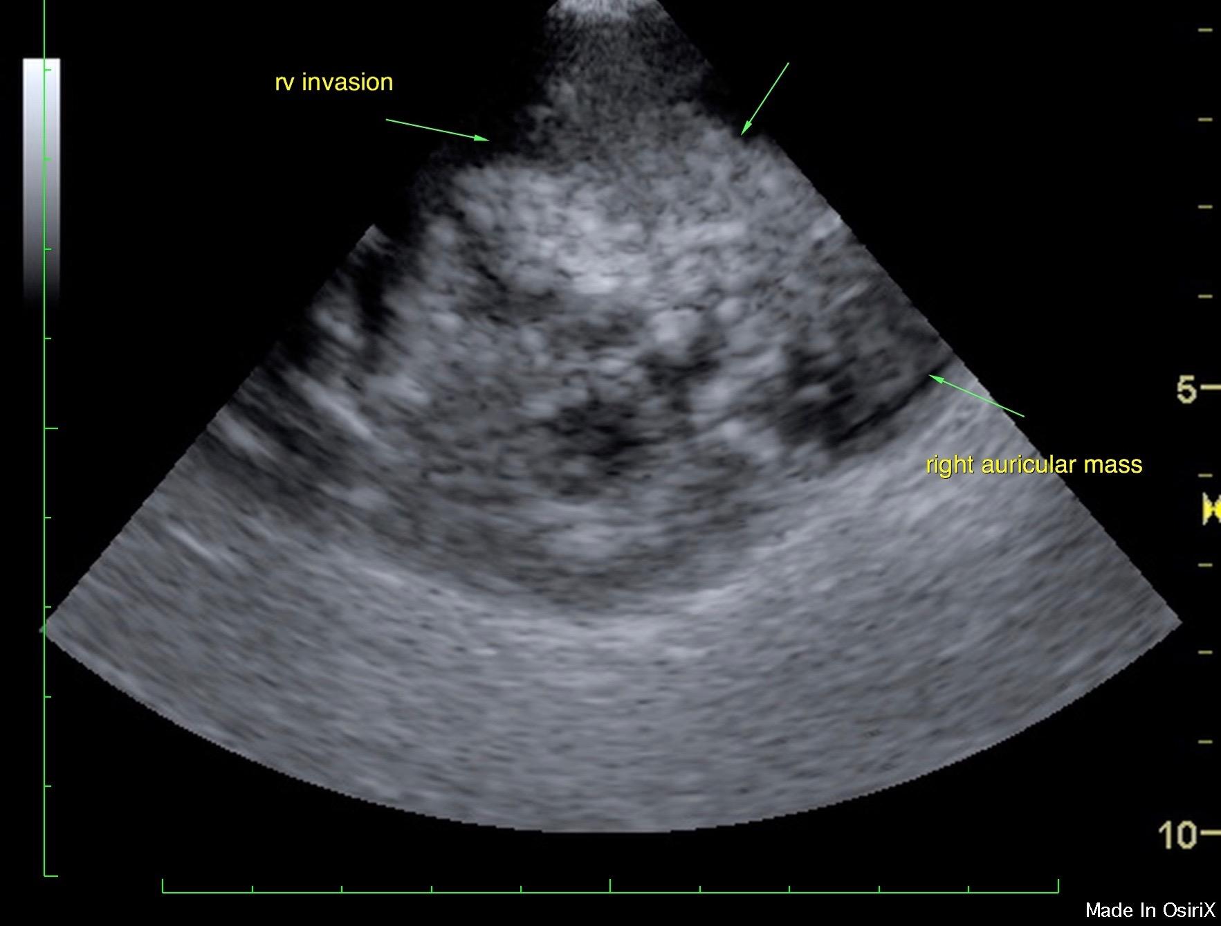
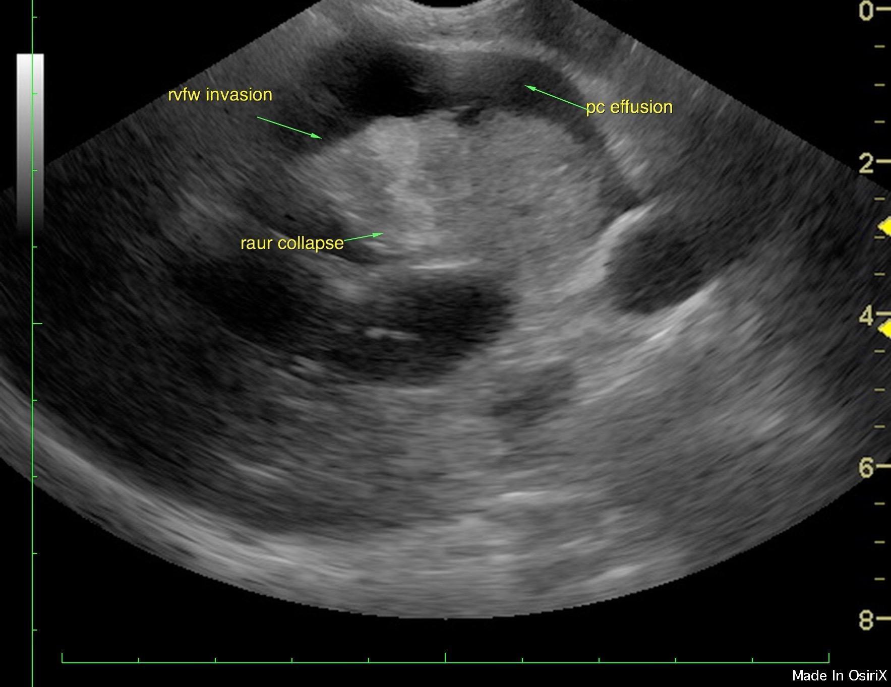
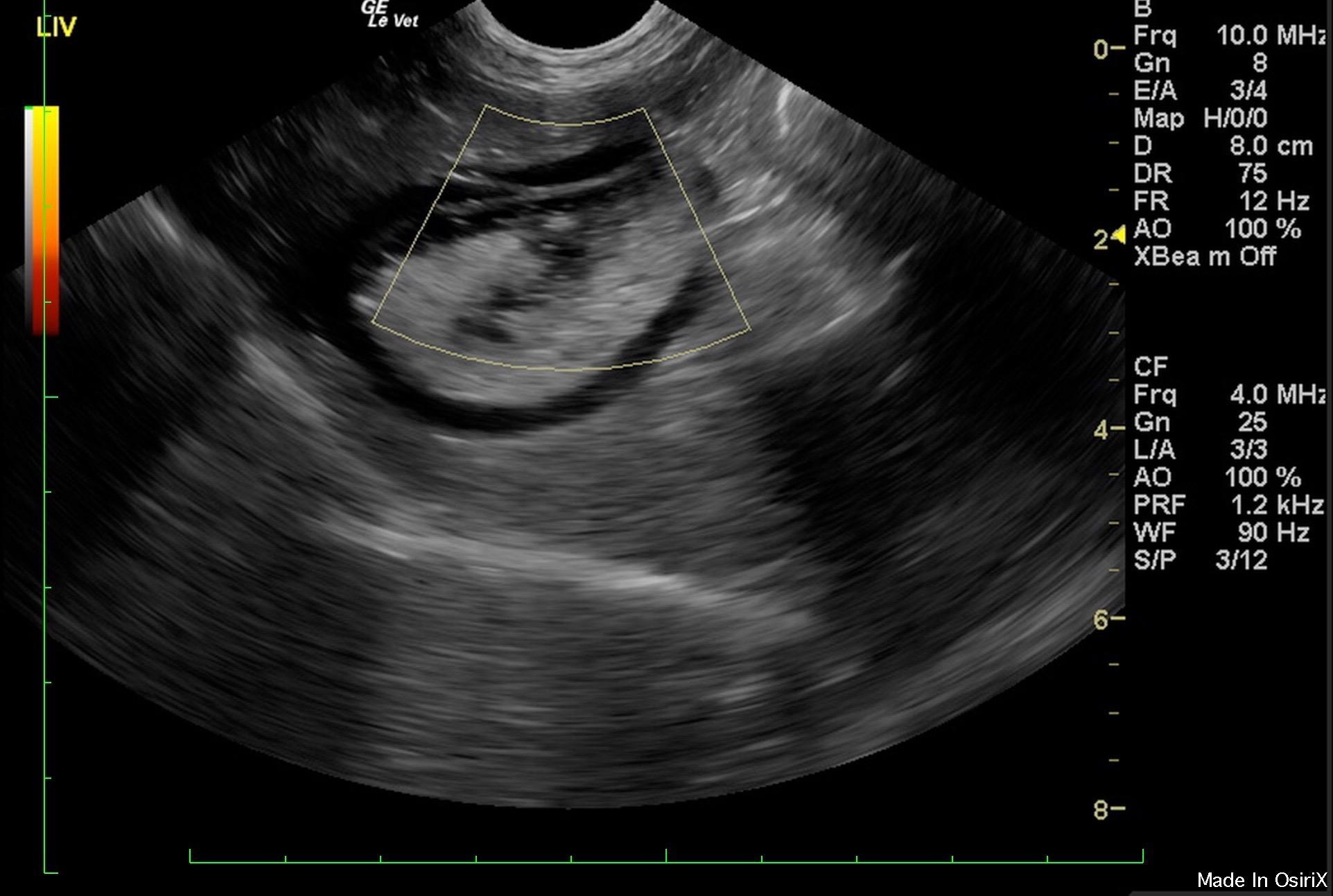
Cardiac presentation revealed a 4 x 3 cm mixed echogenic heart based mass overlying and invading the right heart from the right atrium, tricuspid valve, right ventricle through the pulmonary artery.  Maximum width of the mass measured 3.4 x 3.3 cm on short axis.  The mass appeared to derive from the right auricle which would suggest hemangiosarcoma; however, it invades the right ventricle as well and causes collapse of the right auricle and tamponade effect as well as collapse of the right ventricle.   Moderate amount of pericardial effusion was noted.  Poor volume was noted in the left ventricle.  

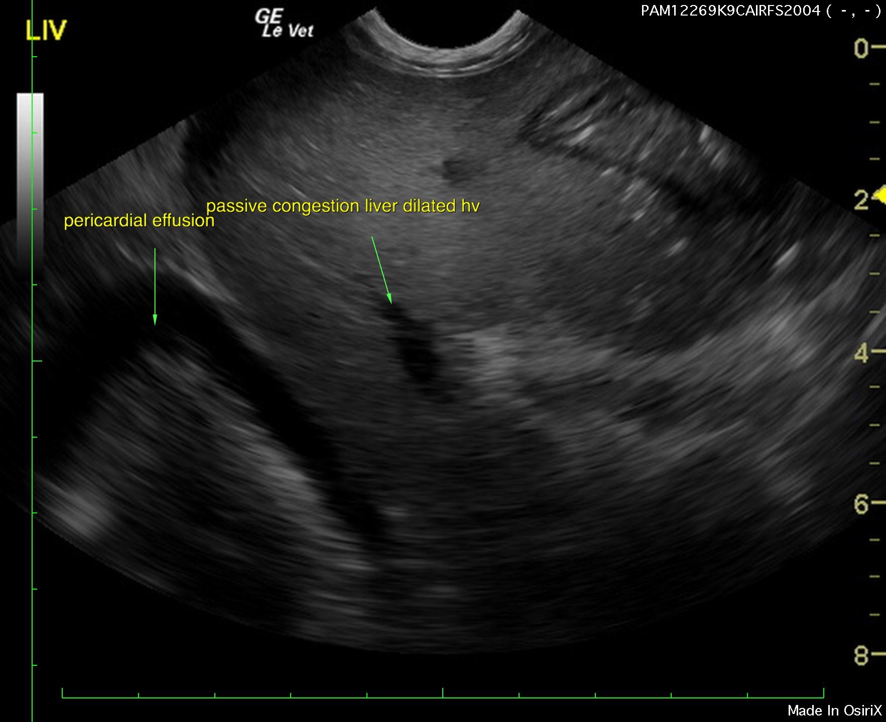
Pericardial effusion was noted in the heart with passive congestion liver pattern and dilated hepatic veins.  
Comments

Pericardiocentesis and potential empirical Adriamycin could be considered in this patient, yet would be based on empirical treatment.
Low-grade chronic inflammatory disease not overtly visible by ultrasound is often detected on FNA or biopsy sample review. Coagulation panel and biopsy may be considered but not considered of immediate necessity unless enzymatic activity (ALT, SAP, Bile acid profile) is elevated. Copper assessment by core biopsy recommended if a chronic ALT/AST component is present in the long term history.

Clinical Differential Diagnosis

Anemia – hemolytic, blood loss
Hypotension – blood loss
Cardiac tamponade
PuPd – renal disease, Cushing’s disease

Patient Information

Patient Name : Lola Debenedicts
Gender : Female, Spayed
Species : Canine
Type of Imaging : Ultrasound
Liz Wuz Here : Yes
Code : 15_00245

Clinical Signs

  • Collapse
  • PU-PD

Images

pam3_1pam1_1pam9_1pam10_1pam6

Blood Chemistry

  • Alkaline Phosphatase (SAP), High

CBC

  • Hematocrit, Low

Clinical Signs

  • Collapse
  • PU-PD
Skip to content