Renal carcinoma vs lymphoma with pleural effusion and hepatic nodules suspicious of metastases in a 14 year old FS DSH cat

Case Study

Renal carcinoma vs lymphoma with pleural effusion and hepatic nodules suspicious of metastases in a 14 year old FS DSH cat

A 14-year-old FS DSH with a history of hyperthyroidism that had been well regulated with the Y/D diet was presented for evaluation weight loss, lethargy, decreased appetite, and intermittent constipation. On physical examination, right-sided renomegaly was present. Serum biochemistry showed elevated BUN (54) and hypercalcemia (3.8).
A 14-year-old FS DSH with a history of hyperthyroidism that had been well regulated with the Y/D diet was presented for evaluation weight loss, lethargy, decreased appetite, and intermittent constipation. On physical examination, right-sided renomegaly was present. Serum biochemistry showed elevated BUN (54) and hypercalcemia (3.8).
DX

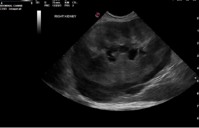
Undefined renal mass. Differentials include carcinoma versus lymphoma or other neoplasia.

Sonographic Differential Diagnosis

Undefined renal mass. Differentials include carcinoma versus lymphoma or other neoplasia.
Regional inflammation.
Noncardiogenic pleural effusion; suspect metastatic disease.
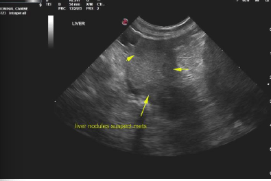
Undefined hepatic nodules; suspicion for metastatic disease.

Image Interpretation

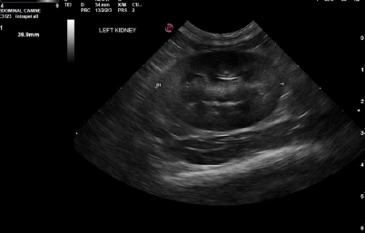
Right kidney in this patient was severely enlarged measuring 8.2 cm with subcapsular hypoechoic halo, disrupted architecture, as well as pyelectasia strongly suggestive for renal carcinoma or possible lymphoma. Pericapsular inflammatory pattern also noted. Left kidney relatively uniform 4 cm, normal size and contour. Corticomedullary definition was maintained.
Minor cystic changes were noted in the liver measuring 1.5 cm most consistent with cystadenoma; this is cranial to the gallbladder in the medial liver adjacent to the diaphragm. This deviated the gallbladder, yet not likely related to the kidney pathology. Subtle hypoechoic nodules were noted in the liver in this patient. Given the renal pathology and the presence of pleural effusion, metastatic disease is a strong potential. Gallbladder was unremarkable. Hepatic veins were not dilated.
Pleural effusion was noted through the diaphragm. Echocardiogram is recommended to assess if this is cardiogenic or related to metastatic disease. Rapid view of the heart revealed volume contraction, which indicates non cardiogenic pleural effusion.

Outcome

Pleurocentesis with Cytospin, FNA of the liver and right kidney are warranted with immediate chemotherapy; however, prognosis is poor.

Clinical Differential Diagnosis

Neoplasia, hydronephrosis, renolith, abscess, granuloma

Patient Information

Patient Name : Prue Anderson/Festival
Gender : Female, Spayed
Species : Feline
Type of Imaging : Ultrasound
Status : Complete
Liz Wuz Here : Yes

Clinical Signs

  • Anorexia
  • Constipation
  • Lethargy
  • Weight loss

History

  • Hyperthyroidism

Images

screen_shot_2016-04-30_at_12screen_shot_2016-04-30_at_12screen_shot_2016-04-30_at_12

Blood Chemistry

  • BUN high
  • Calcium, High

Clinical Signs

  • Anorexia
  • Constipation
  • Lethargy
  • Weight loss
Skip to content