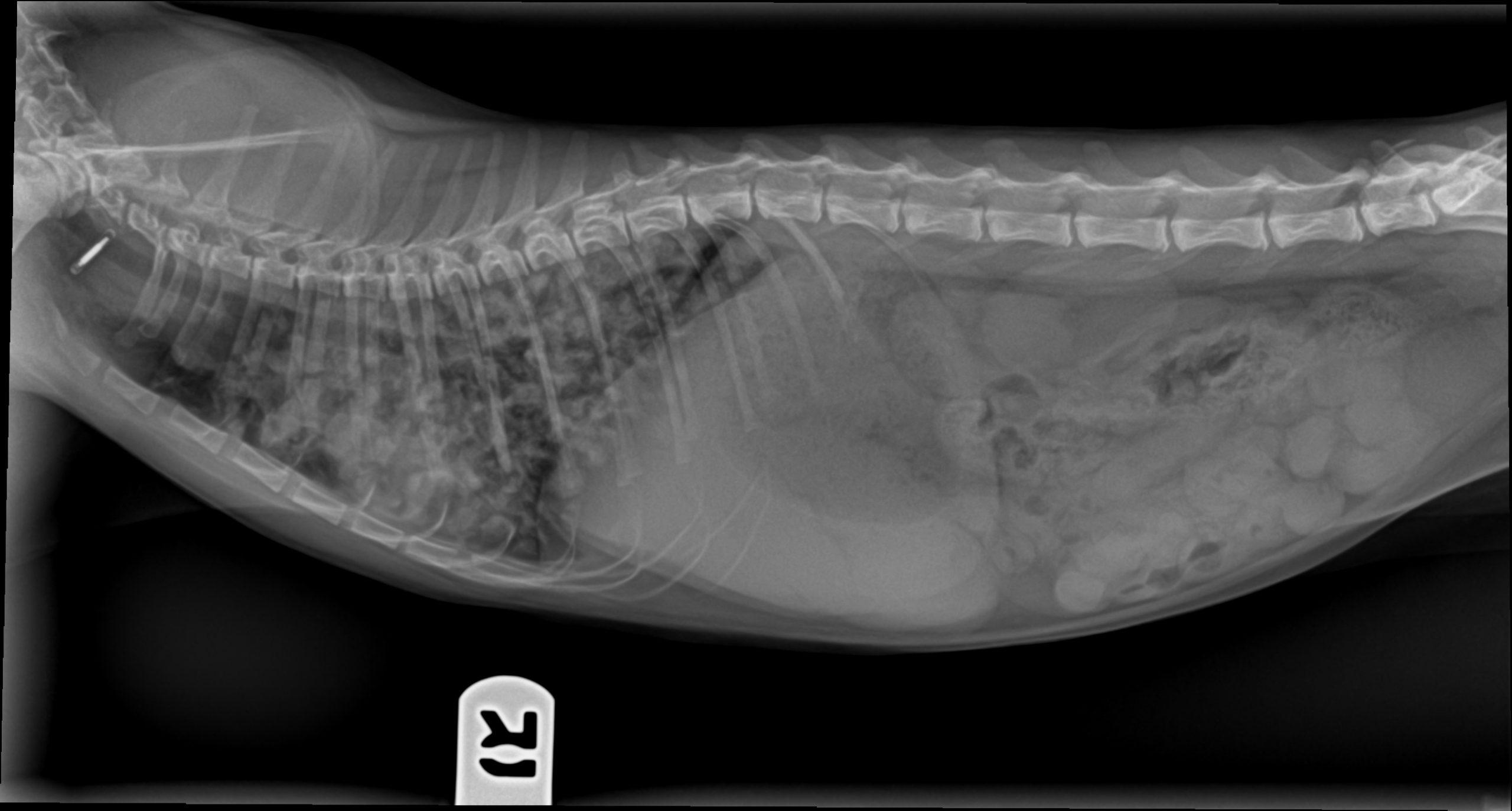
History of respiratory issues (dyspnea, frequent small breaths).
Physical Exam: Normal, mild gingivitis noted. CBC: neutrophils elevated 24,000.
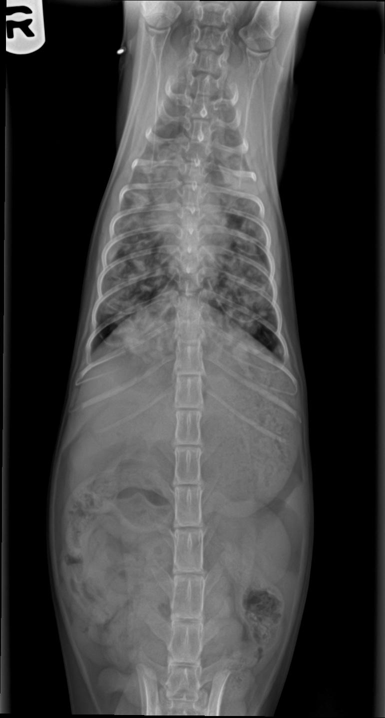
History of respiratory issues (dyspnea, frequent small breaths).
Physical Exam: Normal, mild gingivitis noted. CBC: neutrophils elevated 24,000.