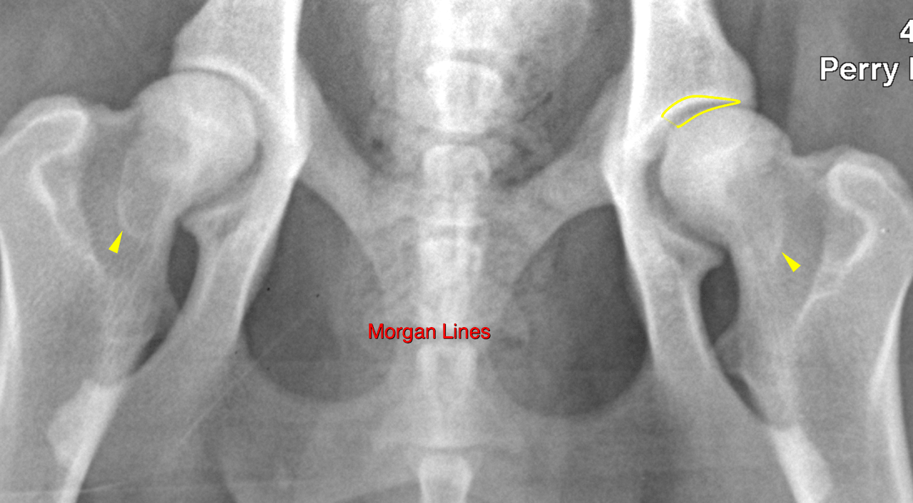
RADS – Minor left-sided hip dysplasia in a 5 year old MN Akita dog

Case Study

RADS – Minor left-sided hip dysplasia in a 5 year old MN Akita dog

This 5 year old MN Akita dog presented with a 2 month history of left hind lameness. Sedated films done at that time showed hip dysplasia with subluxation of left hip. He stands with hind legs base narrow, hocks are together at home. No trauma known.

Physical Exam: base narrow stance in both hind legs. Both stifles thickened medially. No spinal pain. BCS 6/9 Can square sit easily. Under sedation today, CTT and CD esp flexion LH more than RH. No subluxation or ortolani sign appreciated.

CBC/ Chem/UA: wnl.

Reason for Ultrasound Exam: Eval for stifle disease vs hip dysplasia
DX

Minor signs of hip dysplasia on the left side without secondary arthrotic changes.

Image Interpretation

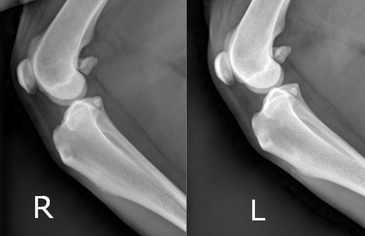
Rads of the stifles, pelvis and lumbar spine –
Right stifle joint:
There is no evidence of increased articular soft tissue opacity. No osteophytes are seen, the periarticular margins are clear. The position of the tibia with respect to the femoral condyles is within normal limits. The popliteal sesamoid bone is in situ.
The tarsal joint is included in the view and is within normal limits.

Left stifle joint:
There is no evidence of increased articular soft tissue opacity. No osteophytes are seen, the periarticular margins are clear. The position of the tibia with respect to the femoral condyles is within normal limits. The popliteal sesamoid bone is in situ.
The tarsal joint is included in the view and is within normal limits.

Pelvis/coxofemoral joints:
There is mild joint space divergence noted for the left coxofemoral joint. A thin Morgan line is seen bilaterally.

Outcome

The radiographic findings do not explain the clinical signs sufficiently.
The sensitivity of radiography is limited for emerging degenerative joint disease and cranial cruciate ligament/meniscal pathology. Consider complementary imaging with superior soft tissue assessment, such as ultrasound, MRI or CT-arthrography for further definition if the clinical suspicion of stifle joint pathology is high despite the negative radiographic exmaination.
Consider muscular/tendinous or spinal disorders as well as underlying causes.

Patient Information

Patient Name : Kane Radford/Perry Hall
Gender : Male, Neutered
Species : Canine
Type of Imaging : Ultrasound
Status : Complete
Liz Wuz Here : Yes

Clinical Signs

  • Lameness

Exam Finding

  • Ambulatory deficits
  • Cranial drawer posiive

Images

bildschirmfoto_2016-04-20_um_15bildschirmfoto_2016-04-20_um_15

Clinical Signs

  • Lameness
Skip to content