RAD study: Probable supraspinatus tendinopathy in a 4-year-old FS Mastiff.

Case Study

RAD study: Probable supraspinatus tendinopathy in a 4-year-old FS Mastiff.

A 4-year-old FS Mastiff mixed breed was presented for limping upon waking on the right front limb. No history of trauma or more activity than normal. The owner reported the patient was lethargic with a decreased appetite. Physical exam found the patient off loading weight from the right front with intermittent lameness, and pain upon flexing and extending right shoulder joint. No swelling or pain upon palpation of long bones or the carpus.
A 4-year-old FS Mastiff mixed breed was presented for limping upon waking on the right front limb. No history of trauma or more activity than normal. The owner reported the patient was lethargic with a decreased appetite. Physical exam found the patient off loading weight from the right front with intermittent lameness, and pain upon flexing and extending right shoulder joint. No swelling or pain upon palpation of long bones or the carpus.
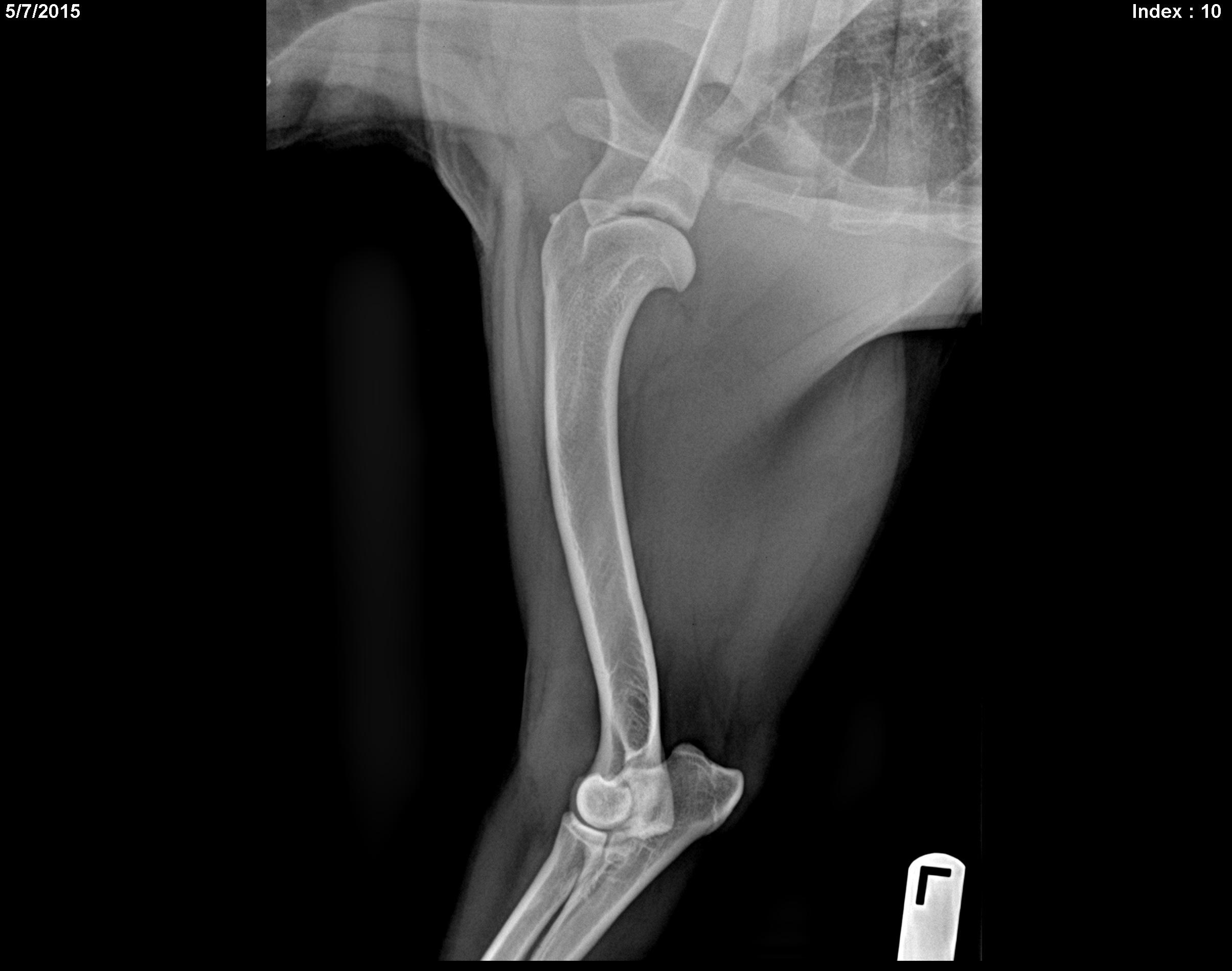
Image Interpretation

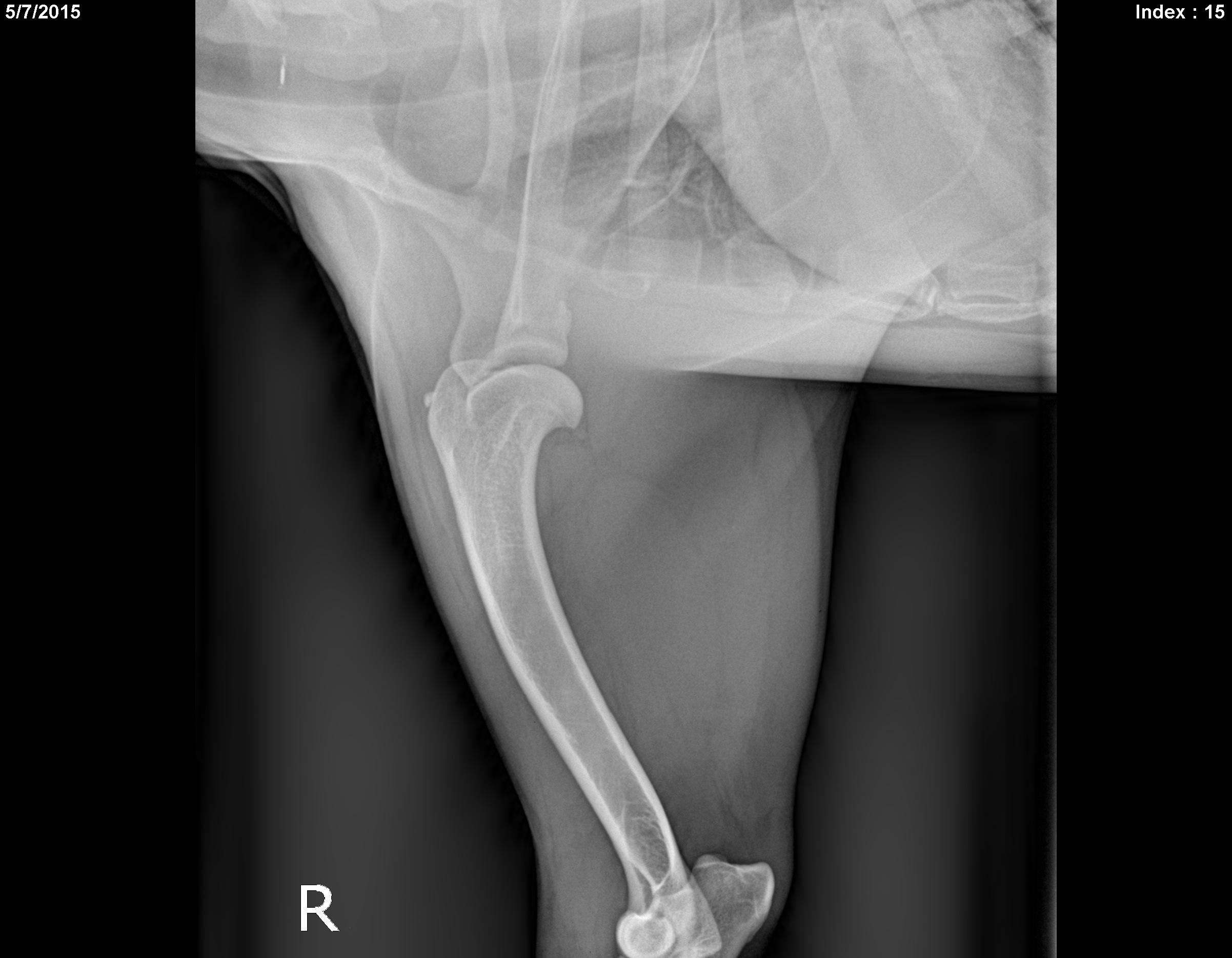
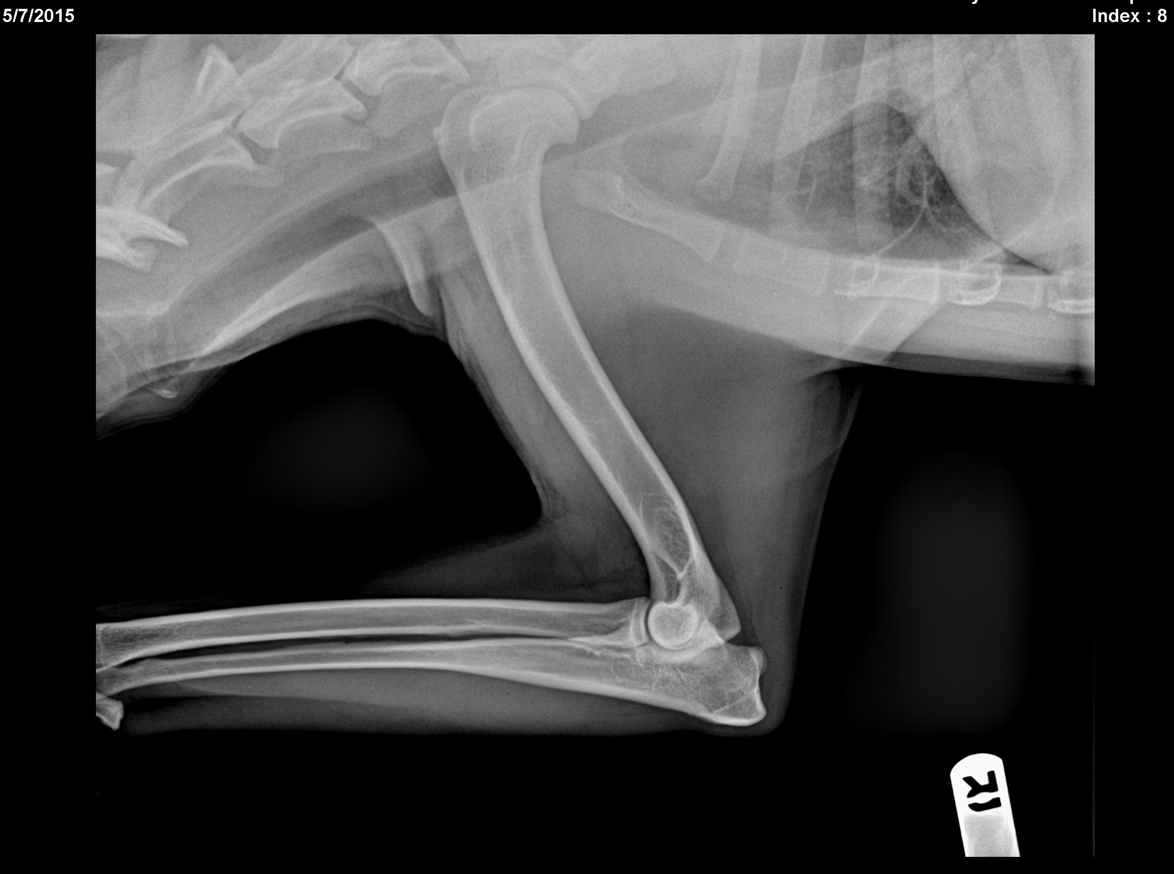
Right Forelimb: A small separate bone opacity (5mm in diameter was seen partially superimposed upon the greater tubercle of the right, proximal humerus. No degenerative changes were seen within
that joint. No aggressive lesions were seen within the humerus or scapula.

Right Elbow: Normal. There was no evidence of degenerative joint disease or other changes.

Right Humerus: Normal. No aggressive changes were noted in any of the bones.

Left Forelimb: A very similar appearing lesion was seen partially superimposed upon the greater
tubercle of the left proximal humerus and measured approximately 3.5 mm. This is the only lesion seen in this leg.

DX

Diagnostic Interpretation: 1. Bilateral, separate bone opacities superimposed upon the greater tubercle - probable supraspinatus tendinopathy ( alternatively , but less likely, bicipital tenosynovitis). 2. Because of the lack of the degenerative joint disease, bicipital tenosynovitis is unlikely. The biceps tendon sheath shares the synovial fluid with the shoulder joint and a chronic degenerative process of the biceps typically leads to degenerative changes noted within the shoulder joint. 3. Lack of such degenerative cha nges and a more cranial position of the separate bone opacities It is much more likely to be associated with the supraspinatus tendon.

Outcome

The recommendations were as follows: Consider should ultrasound bilaterally to further evaluate the supraspinatus tendon and biceps tendons and to better locate this separate bone opacity. Alternatively, consider local injections of anti inflammatories as a diagnostic test and for therapy as clinically indicated. Sometimes there is focal pain on manipulation of the appropriate tendons. The supraspinatus inserts onto the greater tubercle so focal manipulation of the tendon
as it reaches the greater tubercle may be interesting if you illicit local pain response in the right or left shoulders.Consider orthopedic referral or consultation as clinically indicated.

Patient Information

Patient Name : Nyla Devor Perry Hall AH 5/7/15
Gender : Female, Spayed
Species : Canine
Type of Imaging : Ultrasound
Status : Complete

Clinical Signs

  • Difficulty walking
  • Lameness

Images

r_im-0001-0015r_im-0001-0008l_im-0001-0010

Clinical Signs

  • Difficulty walking
  • Lameness