This 5 year old F dog presented with persistent right front lameness that is difficult to localize. 4DX negative.
This 5 year old F dog presented with persistent right front lameness that is difficult to localize. 4DX negative.
This 5 year old F dog presented with persistent right front lameness that is difficult to localize. 4DX negative.
This 5 year old F dog presented with persistent right front lameness that is difficult to localize. 4DX negative.
Rads of the neck, front limbs –
Neck: The facet joints C2/C3 and C3/C4 present emerging osteophyte formation.
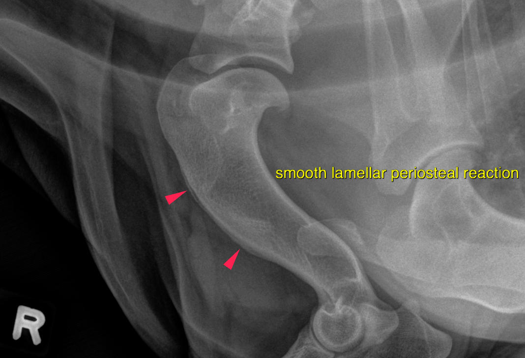
Left front limb: The interosseous ligament/membrane of the radius and ulna presents moderateirregular new bone formation. A triangular bony spur is noted at the lateral aspect of the distal radial metaphysis.
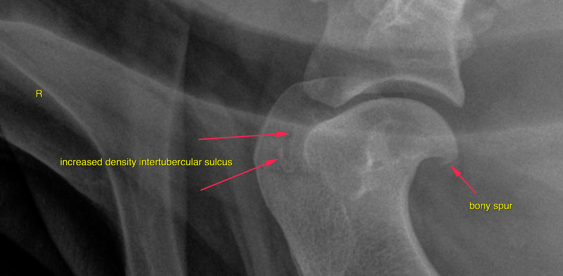
Right front limb: Mild osteophyte new bone formation is noted at the caudal aspect of the humeral head. The intertubercular sulcus presents a mild patchy increased radiopacity. The interosseous ligament/membrane of the radius and ulna presents moderate irregular new bone formation with peripheral sclerosis and a short transition zone to the surrounding bones. A triangular bony spur is noted at the lateral aspect of the distal radial metaphysis.
The possibility of RUIN (radioulnar ischemic necrosis of the interosseous ligament/membrane) should be considered.
Most of these lesions are due to tearing of the bony insertion of the interosseous ligament/interosseous membrane and the severity depends on the involvement of the nutrient blood vessel that enters the ulna within the interosseous attachment. Many of those lesions are incidental, but they carry the potential of causing pain and restricted range of motion.
Even though the dog does not appear to present the typical body type, the findings of
the right shoulder joint is suggestive for tendinopathy of the bicep tendon. Either
ultrasound (can be done consciously in most dogs) or MRI may be considered for
further definition. MRI would offer the option to rule out compressive or other
myelopathy as underlying cause of the lameness at the same time.