RAD – Pulmonary Thromboembolism (PTE) with regional lung infarction and repercussion edema in a 6 year old FS Papillion

Case Study

RAD – Pulmonary Thromboembolism (PTE) with regional lung infarction and repercussion edema in a 6 year old FS Papillion

This 6 year old FS Papillion dog presented with difficulty breathing, increased panting. Rads show enlarged right pulmonary vein.
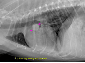
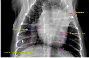
This 6 year old FS Papillion dog presented with difficulty breathing, increased panting. Rads show enlarged right pulmonary vein.

DX

The radiographic changes of the lung support arterial engorgement and hyperperfusion of the lung with a regional interstitial infiltrate and multifocal peripheral hypo perfusion, likely pulmonary thromboembolism (PTE) with regional lung infarction and reperfusion edema

Image Interpretation

left lateral and VD thorax – The caudal compartment of the left cranial lung lobe reveals atelectasis with volume
loss and compensatory midline shift of the heart towards the left. Assessment of the
main pulmonary artery is limited owing to the increase in soft tissue opacity in the left
cranial lung lobe. The right pulmonary artery is dilated, engorged, tortuous in course
and ill defined due to a regional increase in interstitial lung opacity on the VD view.
There is differential perfusion of the caudal lung lobes with a peripheral increase in
interstitial opacity on the right side whereas increased lucency with hardly any
pulmonary vessels can be seen on the left side.
A pleural fissure line is seen between the right middle and right caudal lobes.
Several sites of cylindrical bronchiectasis are seen throughut the lung which are likely
to be an expression of former airway disease and unrelated to the current clinical
presentation.
The heart reveals a mild increase in sternal contact.

Outcome

The changes of the heart are not specific. This case that urgently warrants further workup by
means of a full cardiac echo including assessment for pulmonary hypertension.
Possible underlying causes of PTE include Cushing’s or other systemic syndromes,
Dirofilariosis and other parasitic infections, pancreatitis, protein losing
nephropathy/enteropathy (AT3 loss), pancreatitis, sepsis, DIC, SIRS, systemic
hypertension and other.

Patient Information

Patient Name : Baily Caravela/Hamburg
Gender : Female, Spayed
Species : Canine
Type of Imaging : Ultrasound
Status : Complete
Liz Wuz Here : Yes
Code : 17_00008

Clinical Signs

  • Dyspnea

Exam Finding

  • Panting

Images

screen_shot_2016-05-25_at_4screen_shot_2016-05-25_at_4

Clinical Signs

  • Dyspnea
Skip to content