RAD – Negative metastases check with mild sternal lymph node enlargement in a 8 year old FS Boxer dog with diagnosed cutaneous large cell lymphoma

Case Study

RAD – Negative metastases check with mild sternal lymph node enlargement in a 8 year old FS Boxer dog with diagnosed cutaneous large cell lymphoma

This 8 year old FS Boxer presented for pustules and a welt which over the course of 2 weeks increased in number. FNA/cytology  of 2 of the nodules showed large cell lymphoma
This 8 year old FS Boxer presented for pustules and a welt which over the course of 2 weeks increased in number. FNA/cytology  of 2 of the nodules showed large cell lymphoma
DX

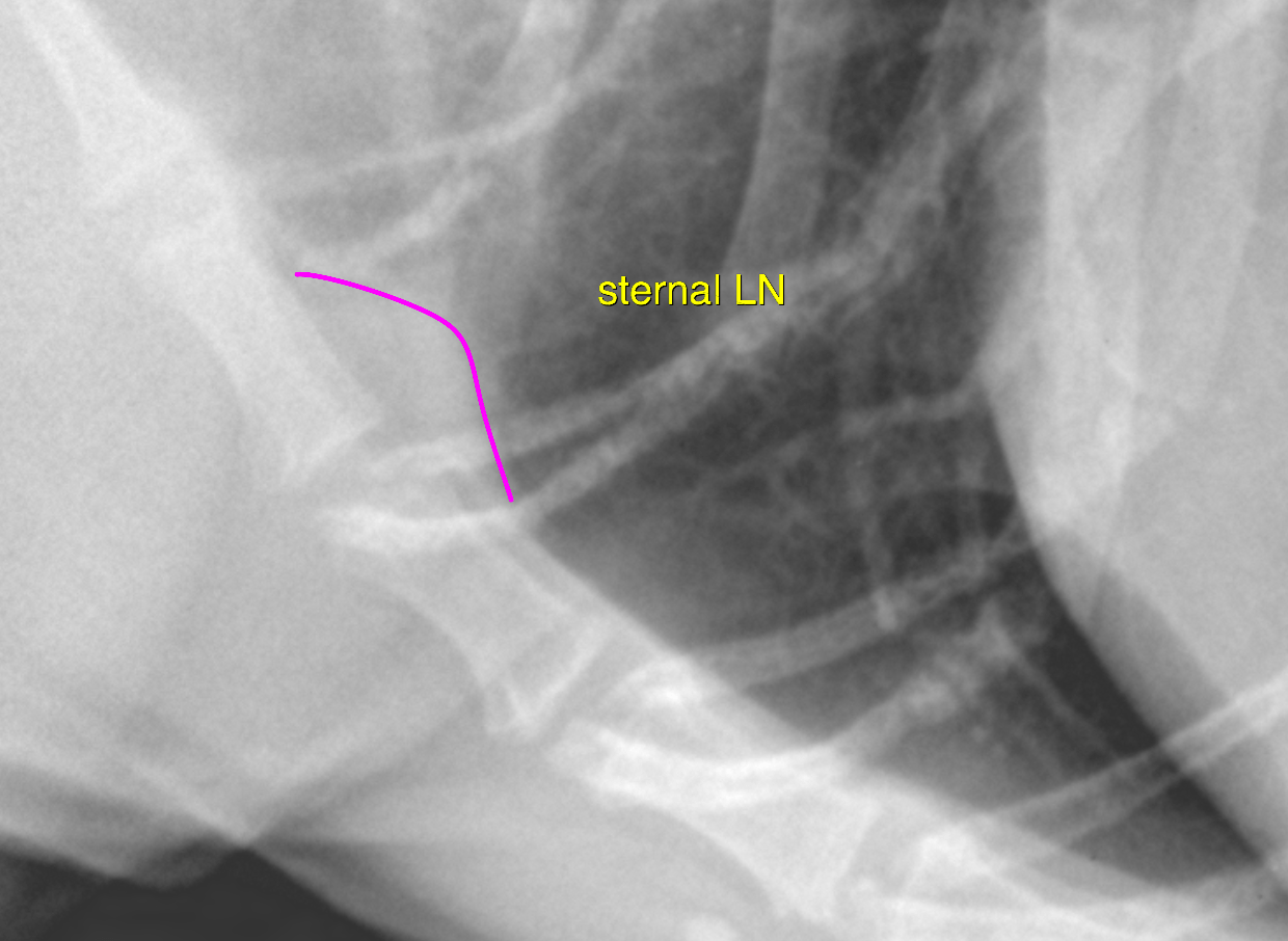
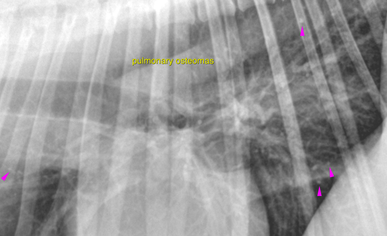
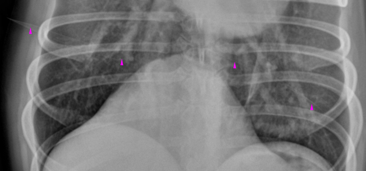
Incidental age related pulmonary osteomas and incidental age related interstitial lung pattern • No evidence of metastatic spread to the lung • Possible mild sternal lymph node enlagement

Image Interpretation

right lateral, left lateral and VD thorax – Multiple, small, irregular
shaped mineral opaque foci are noted throughout the lung parenchyma. Interstitial
nodules are not seen. The lung reveals a mild generalized interstitial pattern. A subtle extrapleural sign is noted on the right- and left-lateral
view level with the sternal lymph nodes.

Outcome

The tributary region for the sternal lymph nodes includes the abdomen, the thoracic
wall and the cranial mammary complexes. The pertaining area should be checked for
possible lesions. A full abdominal ultrasound should be considered owing to the
possible sternal lymphadenomegaly and to complete the tumor staging as such.

Patient Information

Patient Name : Brooke Mungul/PHAH
Gender : Female, Spayed
Species : Canine
Type of Imaging : Ultrasound
Status : Complete
Liz Wuz Here : Yes
Code : 17_00028

Exam Finding

  • Masses

Images

bildschirmfoto_2016-05-24_um_09bildschirmfoto_2016-05-24_um_09bildschirmfoto_2016-05-24_um_09
Skip to content