RAD – Minor esophageal gas dilation in a suspected megaesophagus in a 4 month old F Labrador Retriever

Case Study

RAD – Minor esophageal gas dilation in a suspected megaesophagus in a 4 month old F Labrador Retriever

This 4 month old F Labrador Retriever presented for vomiting; PE transient megaesophagus.

CBC/Chem: HCT 33%, globulin slightly decreased at 2.1
This 4 month old F Labrador Retriever presented for vomiting; PE transient megaesophagus.

CBC/Chem: HCT 33%, globulin slightly decreased at 2.1
DX

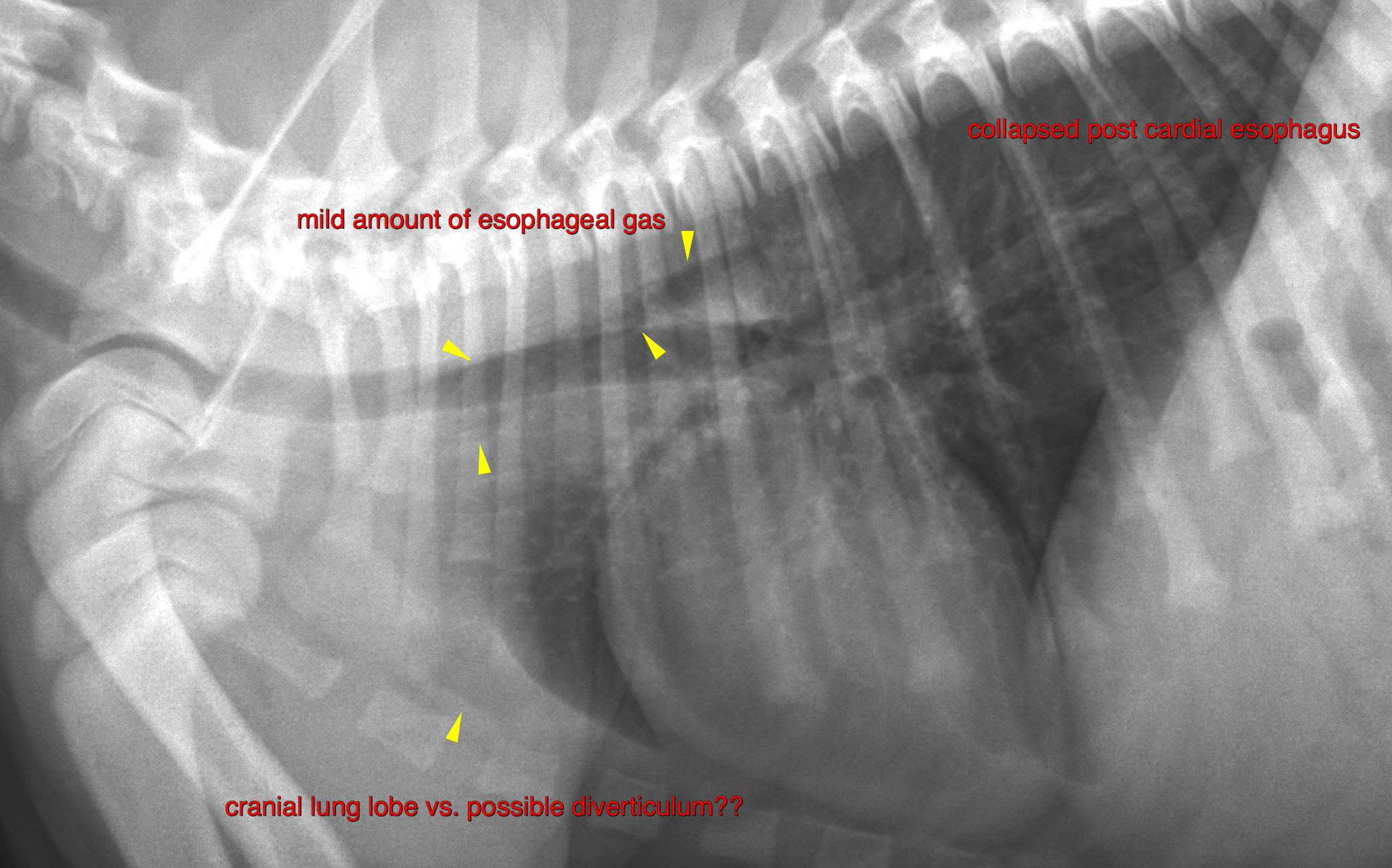
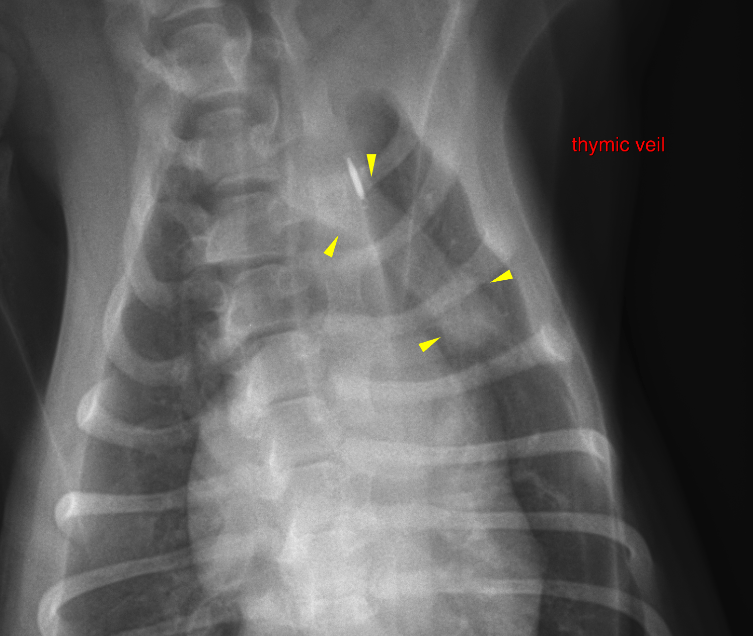
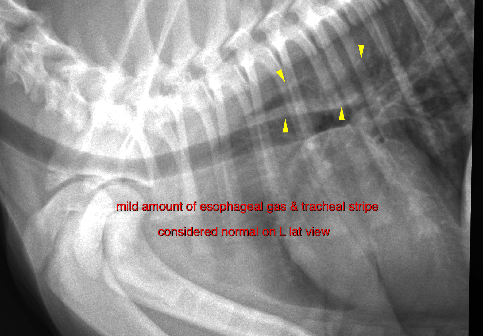
• The radiographs do only show minor segmental dilation of the cranial thoracic esophagus with gas

Image Interpretation

The thymic veil is seen within the cranioventral mediastinum which is an age related normal finding. The esophagus presents an accumulation of a small amount of gas without significant dilation cranial and dorsal to the heart. The cervical esophagus is within normal limits.  A small ovoid retrosternal gas opacity is noted level with the 2nd sternebra on the right lateral view (see picture below).


Outcome

• No evidence of a generalized megaesophagus at the point of the radiographic examination
• The small cranioventral retrosternal gas opacity is likely to consistent with part of the cranial lung lobes. However, the possibility of a cranial thoracic esophageal diverticulum cannot be ruled out entirely.
The radiographs are inconclusive for the presence or cause of a true megaesophagus/esophageal diverticulum. A Barium swallow study is recommended for further definition of the esophageal motility and to rule out a cranioventral diverticulum.

Patient Information

Patient Name : Sophia Ferry/CVC
Gender : Female, Intact
Species : Canine
Type of Imaging : Ultrasound
Status : Complete
Liz Wuz Here : Yes
Code : 17_00065

Clinical Signs

  • Vomiting

Images

bildschirmfoto_2016-08-30_um_13bildschirmfoto_2016-08-30_um_13bildschirmfoto_2016-08-30_um_13

Clinical Signs

  • Vomiting
Skip to content