RAD – Bilateral medial coronoid pathology with flexor and extensor enthesiopathy in a 11 year old MN Chow

Case Study

RAD – Bilateral medial coronoid pathology with flexor and extensor enthesiopathy in a 11 year old MN Chow

This 11 year old Chow mix presented for intermittent lameness of both front limbs.

Physical exam: left front – grade 3/5 lameness, modreate crepitus in carpus, moderate-severe dereased ROM elbow. Hindlimbs – mild crepitus bilateral stiflesm decreased ROM bilateral hips.

This 11 year old Chow mix presented for intermittent lameness of both front limbs.

Physical exam: left front – grade 3/5 lameness, modreate crepitus in carpus, moderate-severe dereased ROM elbow. Hindlimbs – mild crepitus bilateral stiflesm decreased ROM bilateral hips.

DX

• Bilateral medial coronid pathology with flexor and extensor enthesiopathy and moderate secondary osteoarthritis • Emerging degenerative joint disease of the carpometacarpal joints – at this stage this is unlikely to be of clinical significance

Image Interpretation

Rads of the left elbow and carpus –  

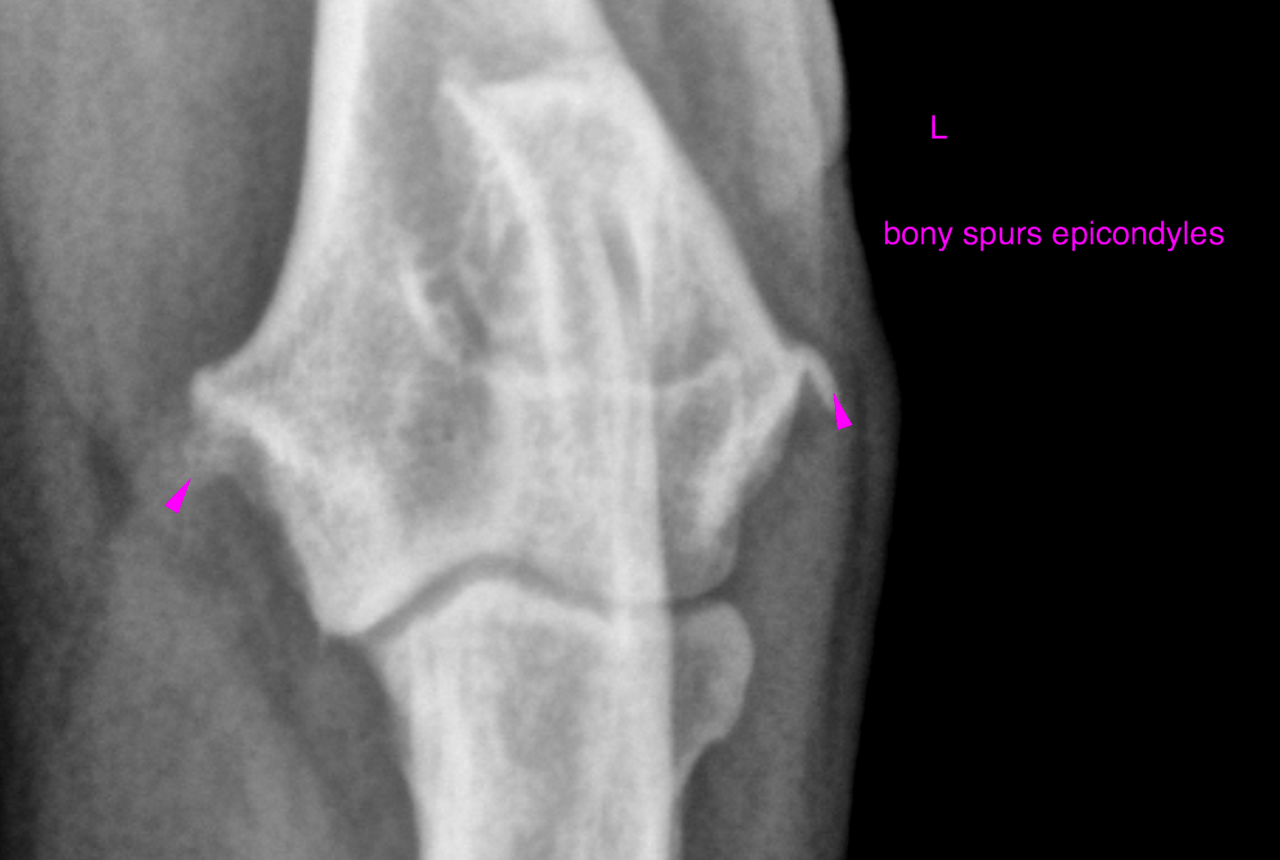
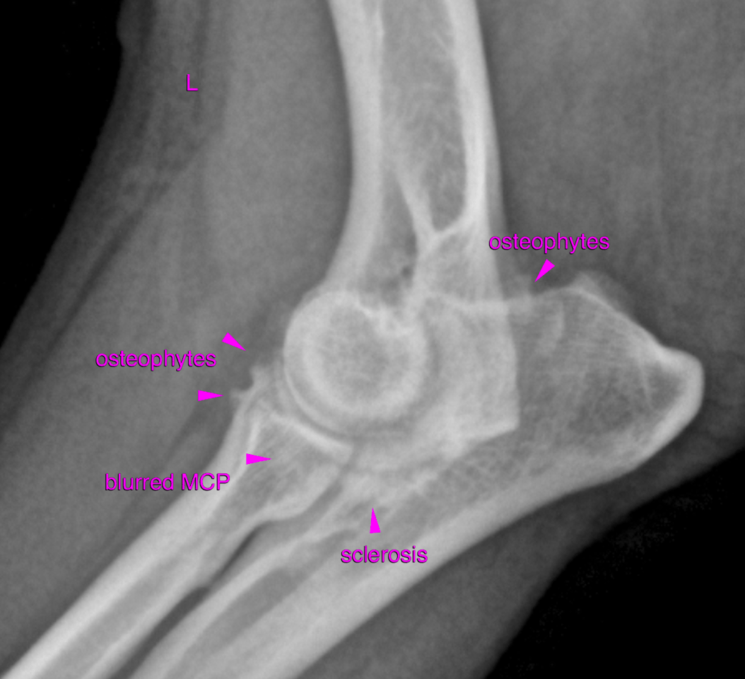
Left elbow- The cranial contour of the medial coronoid process (MCP) is blurred. Marked loss of opacity is noted in the proximal aspect oft he MCP. The trochlear notch of the ulna reveals moderate sclerosis caudal to the MCP. A moderate amount of osteophytes is seen at the pericarticular margins. The medial and lateral epicondylus of the humerus present moderate enthesiophytosis which is an indirect sign of flexor and extensor enthesiopathy. The subhcondral bone of the trochlea humeri is sclerotic but even and smooth with no obvious defects. 

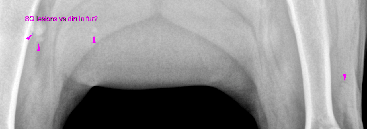
Multifocal small well-delineated soft tissue and mineral opacities are seen superimposed onto the soft tissue (see picture below) and may be dirt in the fur versus soft tissue nodules. Clinical correlation is warranted.

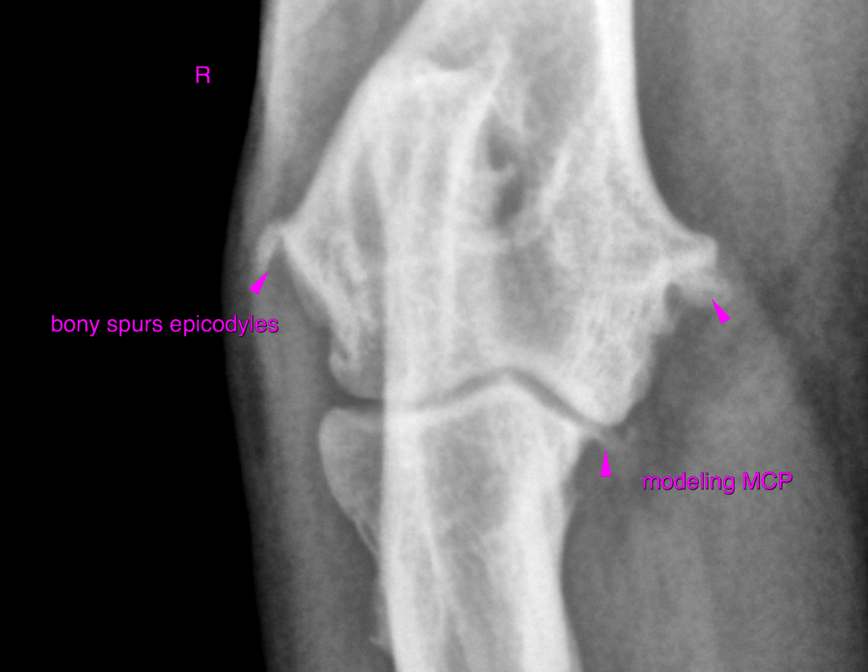
Right elbow – The craniocaudal view of the right elbow presents very similar changes compared with the left one. However the modeling of the MCP is more pronounced in this view.

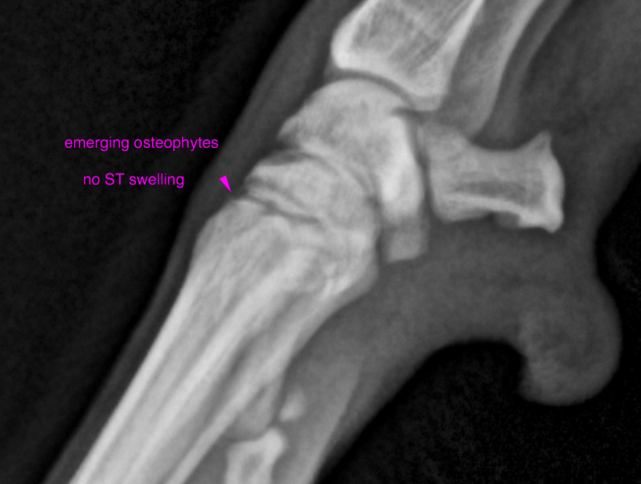
left and right carpus- Very small emerging osteophytes are seen at the periarticular margins of the carpometacarpal joint. There is no evidence of an articular swelling oft he carpal joints.

Outcome

The age of the patient is unusual for the first occurrence of clincial signs linked to medial coronoid pathology only. Late onset elbow dysplasia is typically associated with medial compartment syndrome and/or activated synovialitis. In this case indirect signs of flexor and extensor muscle enthesiopathy are seen as well which may also contribute to the lameness as well. Should the patient be refractory to medical treatment and physical therapy the further management should include pre- and post-contrast CT of both elbows as well as athroscopic revision of the clinically affected joint.

Patient Information

Patient Name : She Hutcheson/DPC
Gender : Male, Neutered
Species : Canine
Type of Imaging : Ultrasound
Status : Complete
Liz Wuz Here : Yes
Code : 17_00041

Clinical Signs

  • Lameness

Images

bildschirmfoto_2016-06-01_um_10bildschirmfoto_2016-06-01_um_10bildschirmfoto_2016-06-01_um_10bildschirmfoto_2016-06-01_um_10bildschirmfoto_2016-06-01_um_10

Clinical Signs

  • Lameness
Skip to content