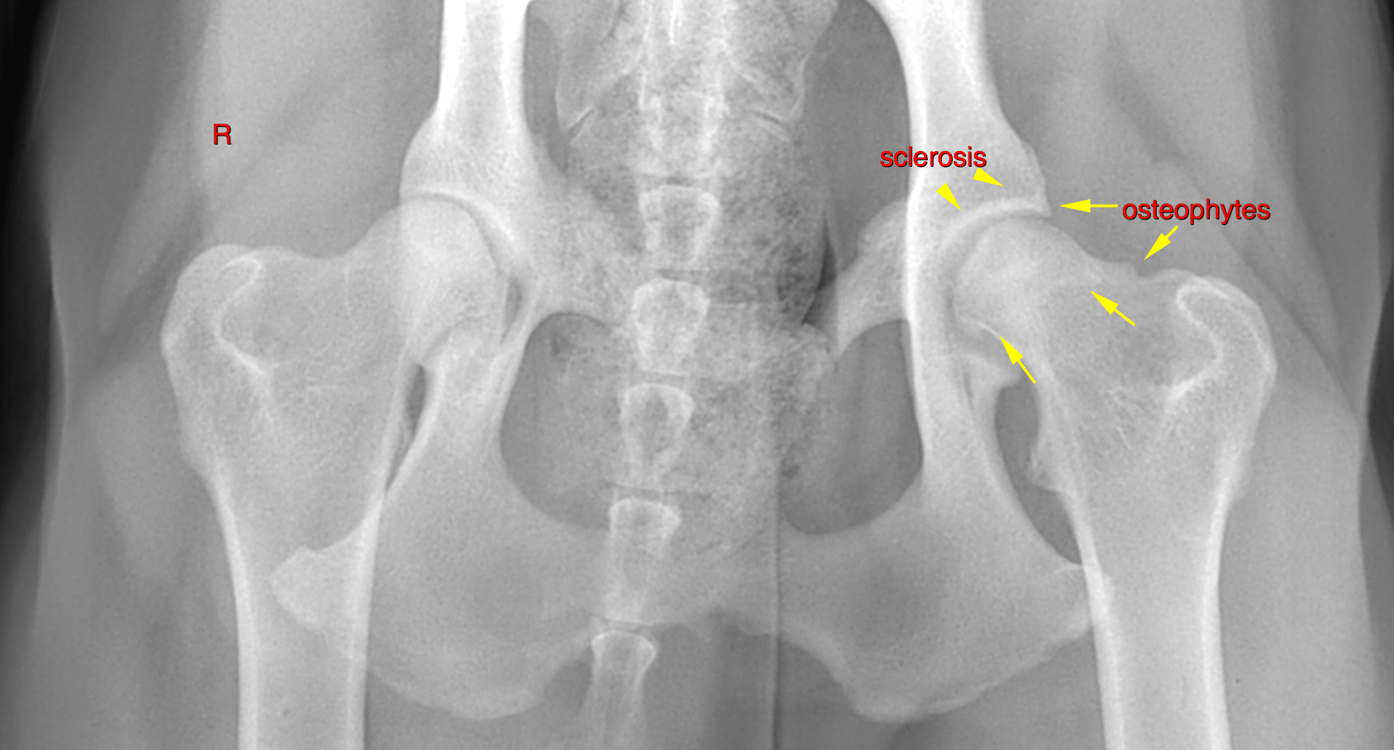
RAD of pelvis/stifles: The left acetabulum presents moderate subchodral bone sclerosis and a mild amount of osteophytes at the acetabular rim. The femoral head and neck present a moderate amount of circumferential osteophytes. Incongruity of the coxofemoral joint space is not noted.
Ultrasound of the stifles:
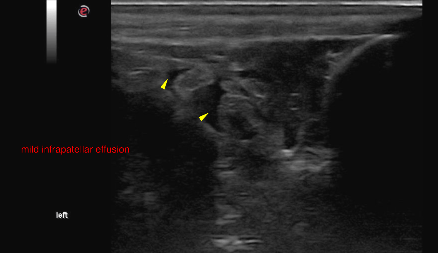
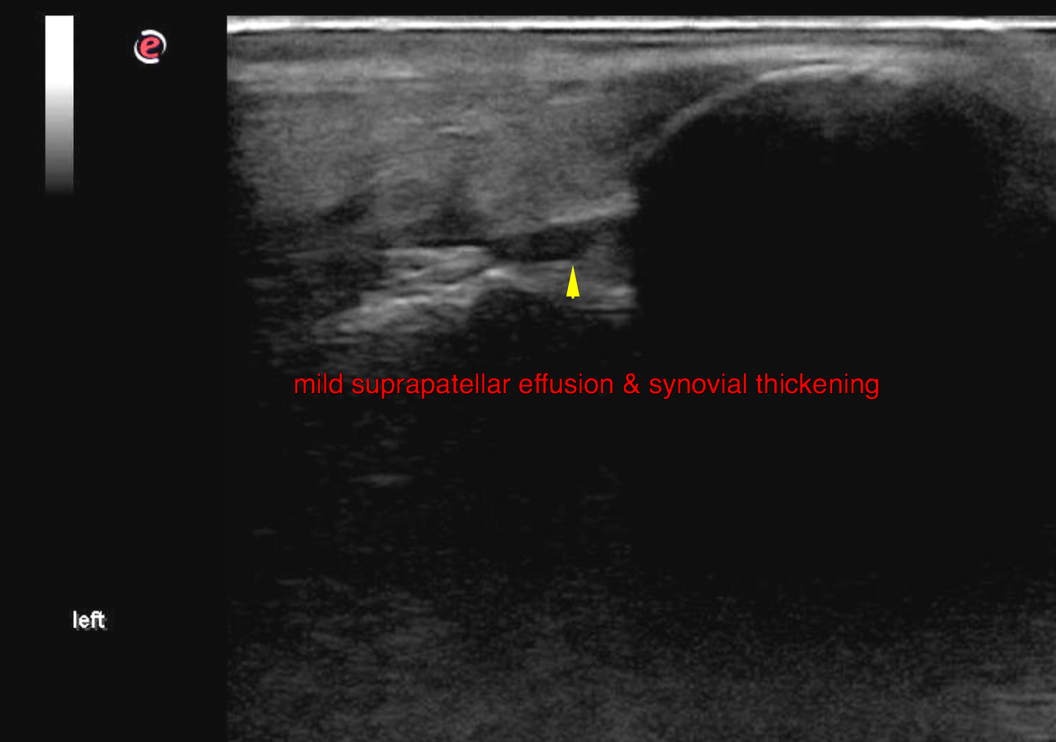
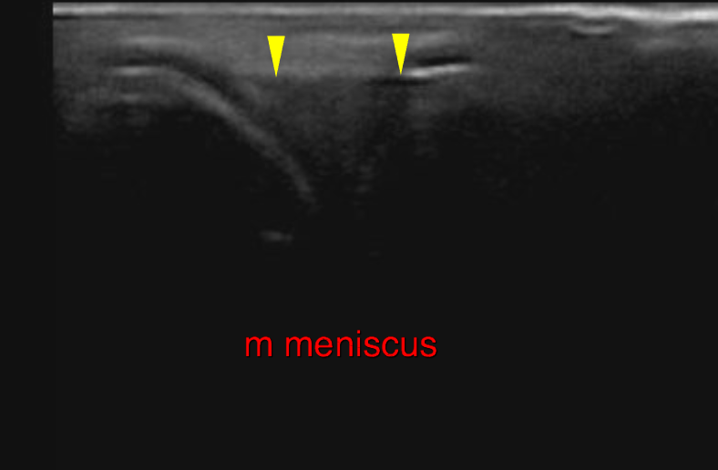
In the left stifle, mild joint effusion is noted within the supra- and infrapatellar recesses. The synovialmembranes present mild thickening emphasizing the area superficial to the cranial cruciate ligament. The cranial cruciate ligament presents continuous with decreased echogenicity, diffuse swelling and slightly undulating margination. The cranial portion of the medial meniscus is seen and within normal limits. The right stifle is similar but with scant joint effusion and the CCL has regular echogenicity and even margination.