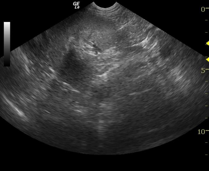
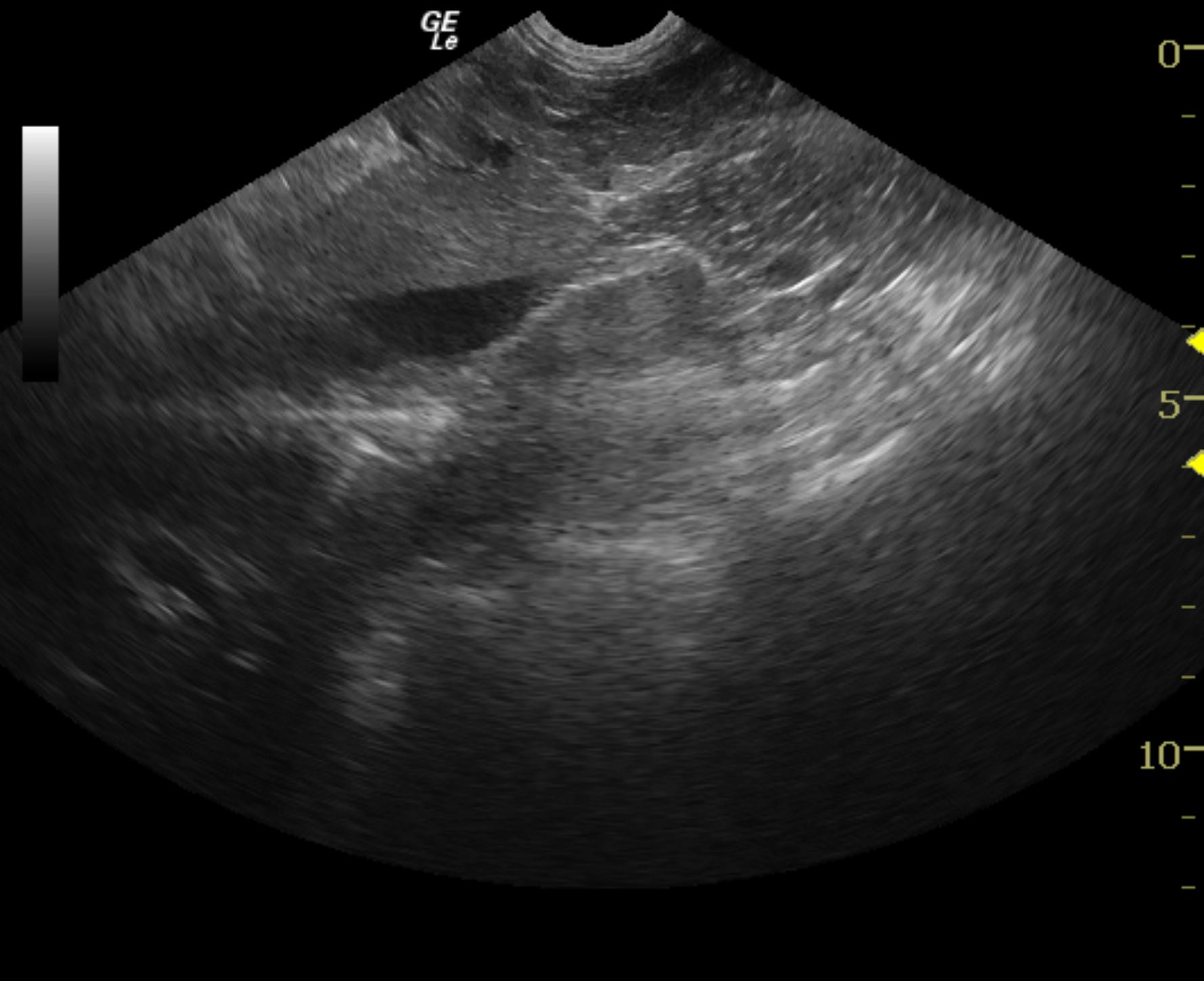
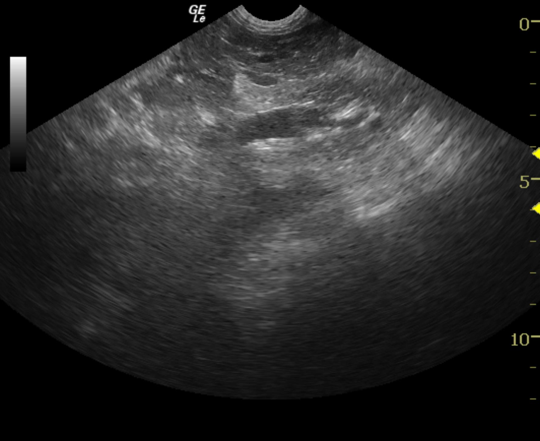
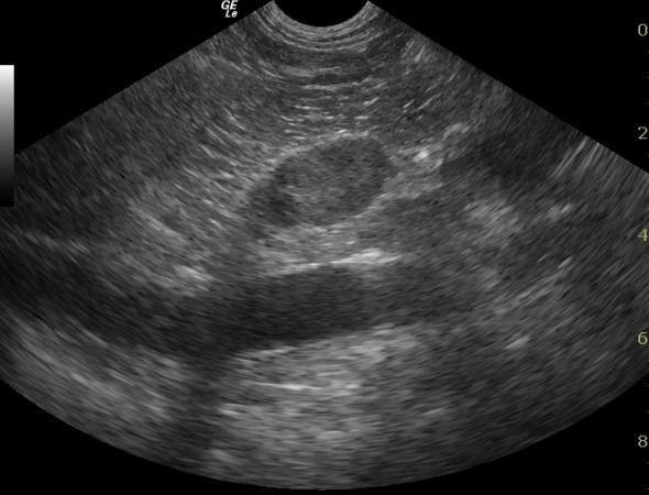
An 8-year-old FS mixed breed dog was referred for partial anorexia and mild lethargy. Mild painful cranial abdomen was noted on physical exam. CBC and blood chemistry panel were unremarkable. Urinalysis revealed 3+ proteinuria with isosthenuria. Systolic blood pressure was 220 mm Hg.
An 8-year-old FS mixed breed dog was referred for partial anorexia and mild lethargy. Mild painful cranial abdomen was noted on physical exam. CBC and blood chemistry panel were unremarkable. Urinalysis revealed 3+ proteinuria with isosthenuria. Systolic blood pressure was 220 mm Hg.