Skip to content
Customize Consent Preferences

We use cookies to help you navigate efficiently and perform certain functions. You will find detailed information about all cookies under each consent category below.

The cookies that are categorized as "Necessary" are stored on your browser as they are essential for enabling the basic functionalities of the site. ... 

Always Active

Necessary cookies are required to enable the basic features of this site, such as providing secure log-in or adjusting your consent preferences. These cookies do not store any personally identifiable data.

No cookies to display.

Functional cookies help perform certain functionalities like sharing the content of the website on social media platforms, collecting feedback, and other third-party features.

No cookies to display.

Analytical cookies are used to understand how visitors interact with the website. These cookies help provide information on metrics such as the number of visitors, bounce rate, traffic source, etc.

No cookies to display.

Performance cookies are used to understand and analyze the key performance indexes of the website which helps in delivering a better user experience for the visitors.

No cookies to display.

Advertisement cookies are used to provide visitors with customized advertisements based on the pages you visited previously and to analyze the effectiveness of the ad campaigns.

No cookies to display.

Parathyroid mass in a 13 year old MN Siberian Husky dog

Case Study

Parathyroid mass in a 13 year old MN Siberian Husky dog

A 13-year-old NM Husky was presented for evaluation of hyperparathyroidism with a serum calcium >16.


DX

Parathyroid enlargement

Sonographic Differential Diagnosis

Left parathyroid adenoma or possible adenocarcinoma with concurrent parathyroid nodule. The presumed adenoma is the internal parathyroid whereas the slightly enlarged nodule is the cranial parathyroid.

Image Interpretation

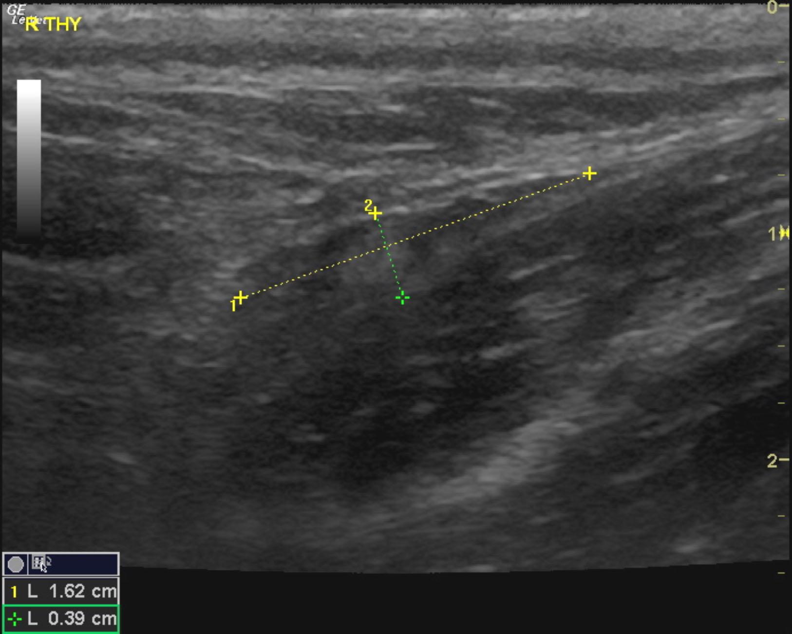
Left thyroid lobe revealed a 1.22 x 0.71 cm nodule with separate hypoechoic nodule cranial to it at 0.3 cm. The thyroid lobe itself measured 3.12 x 0.75 cm. Right thyroid lobe was unremarkable, uniform with hyperechoic nodule consistent with fatty deposit 0.39 cm; right parathyroids not visible. Minor parenchymal remodeling noted. The left internal parathyroid appears to be the enlarged lesion with loss of parenchymal detail and irregular contour most consistent with adenoma, mild potential for parathyroid adenocarcinoma. Cranial to it the 0.29 cm hypoechoic nodule is uniform. No evidence of regional metastasis is noted in the image set provided.

Outcome

Recommendation is removal of both the internal and cranial parathyroid lesions on the left lobe. Full thyroid panel is recommended in this patient if T4 is subnormal.

Comments

Patient lost to follow-up

Clinical Differential Diagnosis

Parathyroid – hyperplasia, adenoma, carcinoma
Toxicity – calcium containing rodenticide

Sampling

None

Patient Information

Patient Name : Coco M
Gender : Male, Neutered
Species : Canine
Type of Imaging : Ultrasound
Status : Complete
Liz Wuz Here : Yes
Code : 13_00039

Images

coco_martinez_parathyroid_adenoma_3coco_martinez_parathyroid_adenoma_2coco_martinez_parathyroid_adenomacoco_martinez_parathyroid_adenoma_rt_thy_normal_fat_deposit

Blood Chemistry

  • Calcium, High