A 6-year-old neutered male Yorkshire terrier mixed breed dog was initially presented at an emergency referral facility for vomiting and diarrhea. Laboratory testing showed abnormal Snap cPLI, decreased BUN (5), hypokalemia, PCV of 43% and TS at 7 g/dL. Survey radiographs had shown a small amount of gas in the intestines and mild hepatomegaly. A diagnosis of pancreatitis was made and the dog treated with SQ fluids, Pepcid, Cerenia, Baytril, Buprenex and fed a bland diet, which resulted in a marked improvement.
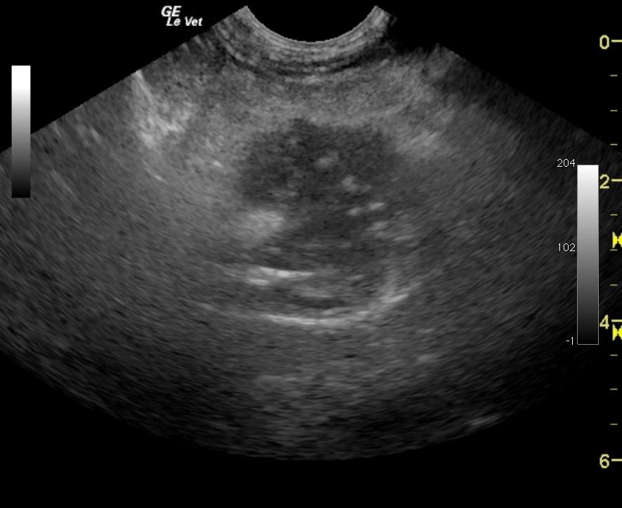
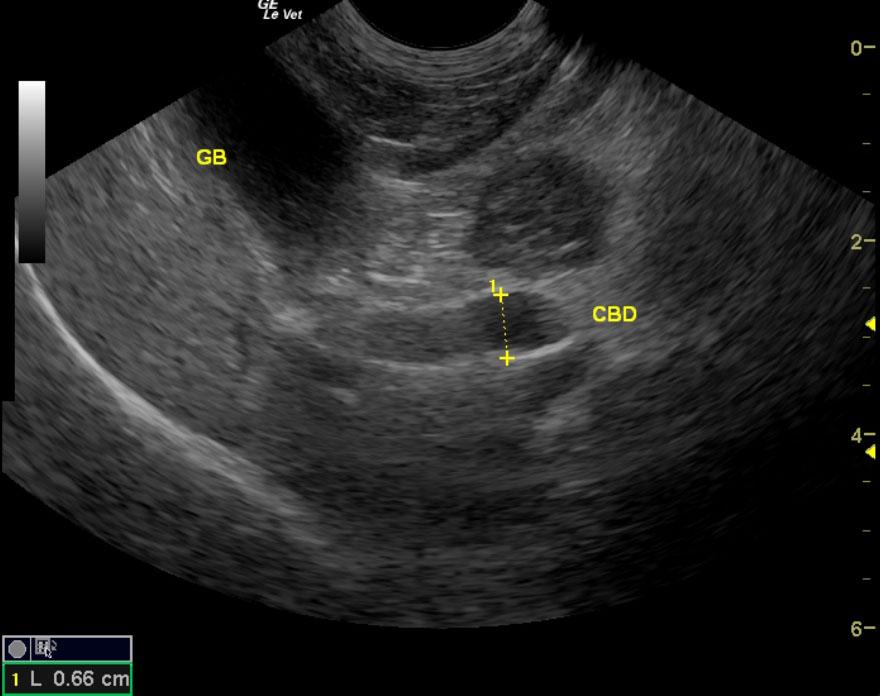
A 6-year-old neutered male Yorkshire terrier mixed breed dog was initially presented at an emergency referral facility for vomiting and diarrhea. Laboratory testing showed abnormal Snap cPLI, decreased BUN (5), hypokalemia, PCV of 43% and TS at 7 g/dL. Survey radiographs had shown a small amount of gas in the intestines and mild hepatomegaly. A diagnosis of pancreatitis was made and the dog treated with SQ fluids, Pepcid, Cerenia, Baytril, Buprenex and fed a bland diet, which resulted in a marked improvement. Two weeks after treatment, the pet ingested a piece of pizza resulting in episodes of vomiting and diarrhea. The pet was treated with Cerenia, SQ fluids, Buprenex, and Baytril to which there was a transient response. The patient was then re-evaluated for vomiting, depression, weakness, and anorexia. On physical examination, the abdomen was tense on palpation, and the mucous membranes were icteric. Serum chemistry showed elevated ALP (> 2000) and elevated ALT (304) activity.