ORTHO – Rupture of the left cranial cruciate ligament (CCL) with degenerative joint disease (DJD) in a 6 year old FS Weimaraner dog

Case Study

ORTHO – Rupture of the left cranial cruciate ligament (CCL) with degenerative joint disease (DJD) in a 6 year old FS Weimaraner dog

The patient is a 6 year old FS Weimeraner dog presented for R/O of a cruciate meniscus tear in the left stifle. Lame on left hind. 

The patient is a 6 year old FS Weimeraner dog presented for R/O of a cruciate meniscus tear in the left stifle. Lame on left hind. 

Image Interpretation

Ultrasound of the left and right stifles – Left stifle joint: 
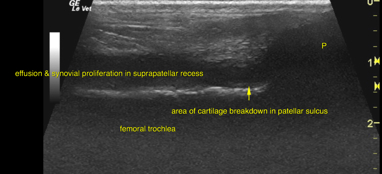
Both the supra- and infrapatellar recess of the left stifle joint contain a moderate
amount of effusion. The joint capsule reveals moderate thickening. Moderate synovial
proliferations are noted.
The cartilage layer as appreciated within the patellar sulcus and onto the femoral
condyles presents with multifocal areas of uneven thickness and outline with increase
of the cartilage’s echogenicity. The subchondral bone of the femoral condyles as
appreciated is slightly uneven in outline.
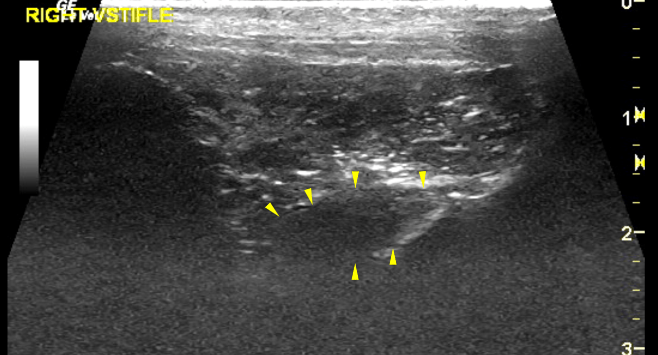
A severe amount of osteophytes is noted at the periarticular margins of the
femoropatellar and femorotibial joints.
The cranial cruciate ligament (CCL) presents as a short stump of echogenic fibres next
to its distal insertion at the intercondylar eminence of the tibia. There is a moderate
amount of periligamentous effusion noted.
The infrapatellar fat body is heterogenous and contains cystic changes as found
commonly in degenerative joint disease (DJD). 
The medial meniscus is small, irregular in outline and presents multifocal increase in
echogenicity. The position of the medial meniscus is in situ. There is perimeniscal
effusion with proliferations of reactive tissue in the meniscal region.
Right stifle joint: 
Both the supra- and infrapatellar recess of the right stifle joint contain a mild amount
of effusion. The joint capsule reveals mild thickening with mild synovial
proliferations.
The cartilage layer as appreciated within the patellar sulcus and onto the femoral
condyles presents with even thickness and surface. The cartilage is anechoic as
expected. The subchondral bone of the femoral condyles as appreciated is even in
outline.
A mild amount of osteophytes is noted at the periarticular margins of the
femoropatellar and femorotibial joints.
The cranial cruciate ligament (CCL) is midly thickened and increased in echogenicity.
The outline of the CCL reveals mild surface irregularity. Except for small focal
hyperechoic areas the fibre pattern is largely maintained. No periligamentous effusion
is seen.
The medial meniscus is in situ with mildly uneven surface and uniform echogenicity.
No superficial tearing is seen. There is no perimeniscal effusion.

DX

The ultrasonographic findings of the left stifle joint are compatible with a complete rupture of the cranial cruciate ligament. The findings of the right stifle joint are compatible with mild generalized chronic degenerative joint disease with joint effusion, osteophytosis, and capsular fibrosis

Outcome

The left stifle joint is affected by a severe
generalized chronic degenerative joint disease with joint effusion, multifocal cartilage
breakdown, osteophytosis, synovial proliferation and capsular fibrosis. Moreover
medial meniscal damage is suggested by the ultrasonographic findings. Generalized
degeneration and disruption by repetitive mechanical trauma is more likely than an
actual tear with the current presentation.
The
right cranial cruciate ligament presents signs of advanced degeneration and edema.
Emerging fibre disruption is a potential but a partial or complete rupture is not overtly
seen and biomechanical failure is not likely at this point.
Surgical therapy is indicated for the left stifle joint to address the biomechanical
instability and prevent progress of the degeneration joint & damage.
Patient Information

Patient Name : Ingrid Hirschenson, Morris Hills
Gender : Female, Spayed
Species : Canine
Type of Imaging : Ultrasound
Status : Complete
Liz Wuz Here : Yes

Clinical Signs

  • Lameness

Images

bildschirmfoto_2016-01-08_um_10bildschirmfoto_2016-01-08_um_10bildschirmfoto_2016-01-08_um_10bildschirmfoto_2016-01-08_um_10bildschirmfoto_2016-01-08_um_11bildschirmfoto_2016-01-08_um_11bildschirmfoto_2016-01-08_um_11

Clinical Signs

  • Lameness