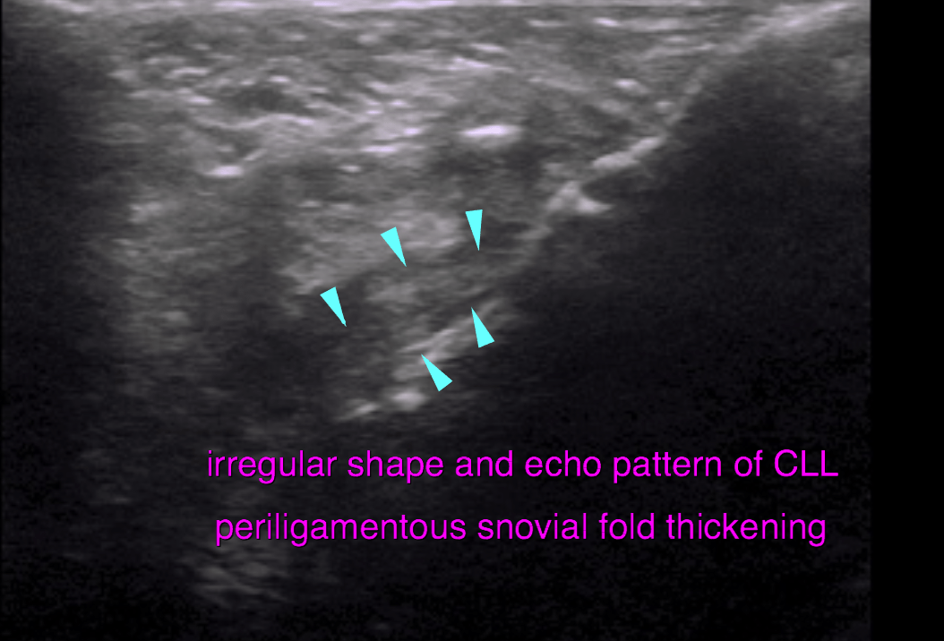
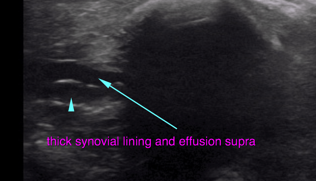
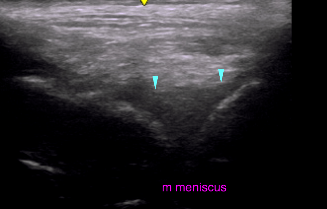
This 4 year old MN Chesapeake Bay Retriever presented for lameness right hind; positive cranial drawer and CTT, suspect partial CCL tear.
This 4 year old MN Chesapeake Bay Retriever presented for lameness right hind; positive cranial drawer and CTT, suspect partial CCL tear.