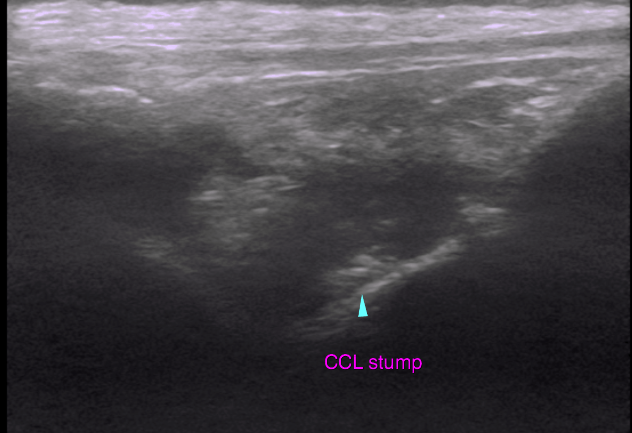
This 5 year old FS Labrador Retriever had previous TTA surgery for ruptured left CCL 6 months ago. Presented with right hind lameness. Positive cranial drawer, CTT right stifle.
This 5 year old FS Labrador Retriever had previous TTA surgery for ruptured left CCL 6 months ago. Presented with right hind lameness. Positive cranial drawer, CTT right stifle.