ORTHO – Mild generalized degenerative joint disease (DJD) with synovialitis, effusion, osteophytosis and partial CCL rupture in a 2 year old MN Newfoundland

Case Study

ORTHO – Mild generalized degenerative joint disease (DJD) with synovialitis, effusion, osteophytosis and partial CCL rupture in a 2 year old MN Newfoundland

This 2 year old MN Newfoundland dog presented with sudden onset left hind lameness. Radiography suggests avulsion fracture of the insertion of the CCL

This 2 year old MN Newfoundland dog presented with sudden onset left hind lameness. Radiography suggests avulsion fracture of the insertion of the CCL

DX

The findings of the left stifle joint are compatible with moderate generalized chronic degenerative joint disease (DJD) with synovialitis as well as moderate joint effusion and mild osteophytosis

Image Interpretation

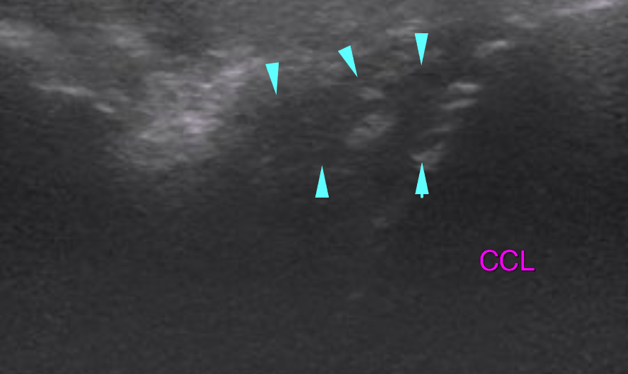
Ultrasound of the left stifle – 

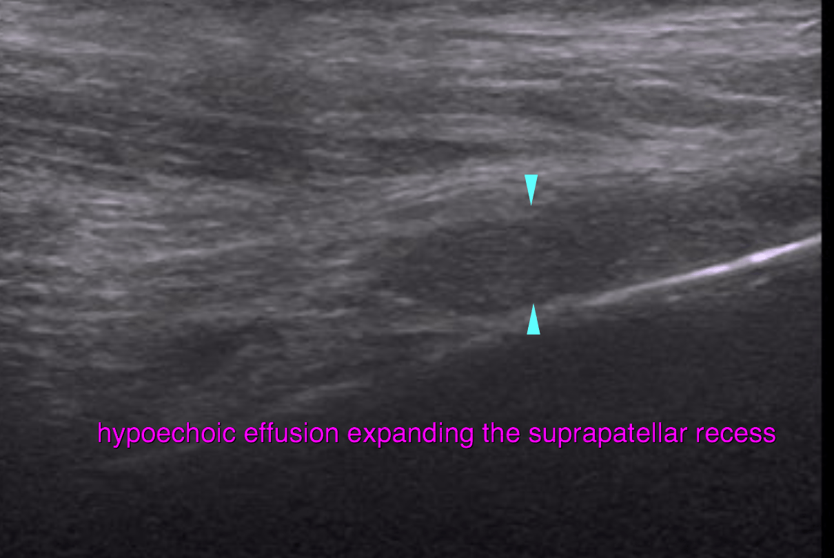
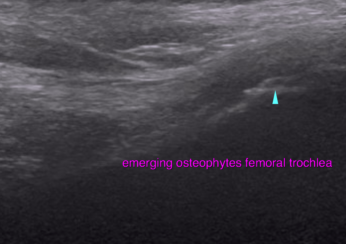
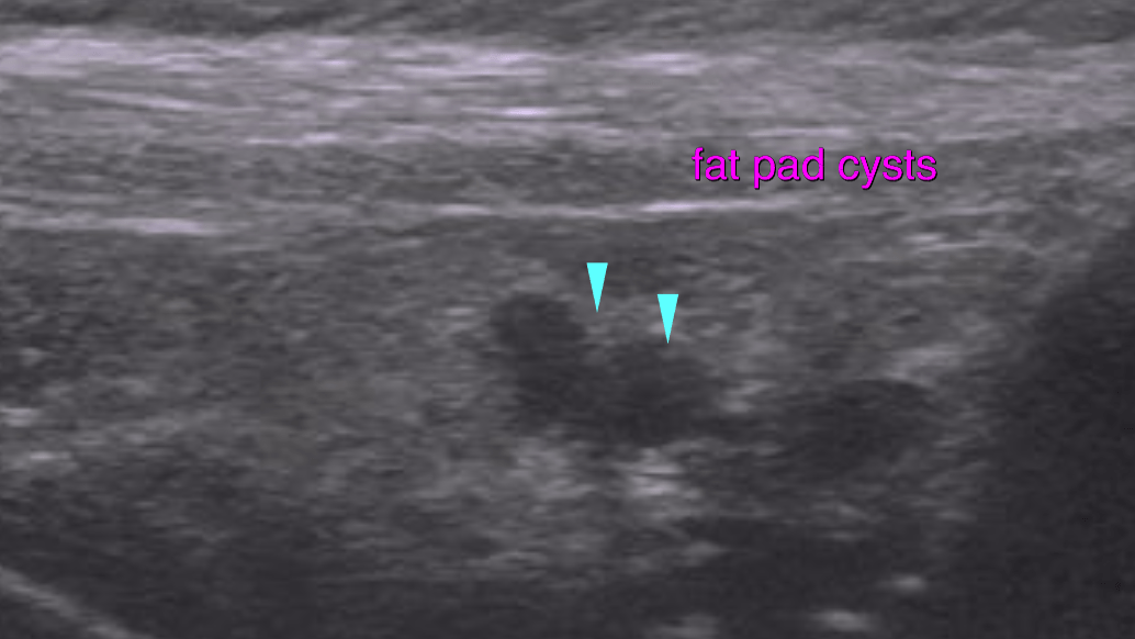
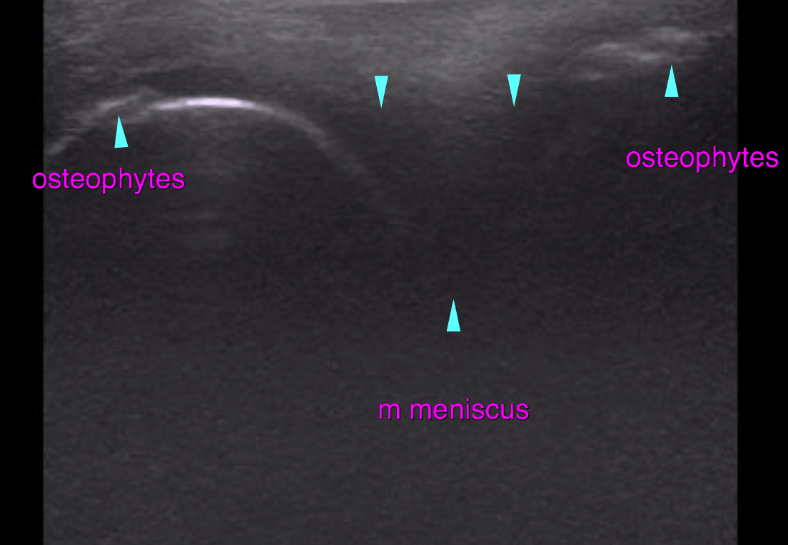
The supra- and infrapatellar recess of the left stifle joint reveal moderate synovial thickening as well as moderate anechoic effusion. A mild to moderate amount of osteophytes is seen at the periarticular margins of the femoropatellar and femorotibial joint. The cranial cruciate ligament (CCL) presents irregular outline, thickness and echogenicity. A mineralized focus is seen within the infrapatellar recess next to the CCL. Mild periligamentous effusion and regional synovial membrane thickening are seen. The infrapatellar fat body presents mild heterogeneity as found commonly in degenerative joint disease (DJD). 

Outcome

There findings are suggestive for partial partial rupture of the cranial cruciate ligament. Even though a traumatic avulsion fracture cannot be ruled out here the overall changes are as typical in degenerative joint disease. I do consider early onset of DJD as underlying pathology here more likely than a strictly traumatic injury.

Patient Information

Patient Name : Odin Alessi/Ridgewood
Gender : Male, Neutered
Species : Canine
Type of Imaging : Ultrasound
Status : Complete
Liz Wuz Here : Yes
Code : 18_00019

Clinical Signs

  • Lameness

Images

bildschirmfoto_2016-06-16_um_18bildschirmfoto_2016-06-16_um_18bildschirmfoto_2016-06-16_um_18bildschirmfoto_2016-06-16_um_18bildschirmfoto_2016-06-16_um_18

Clinical Signs

  • Lameness
Skip to content