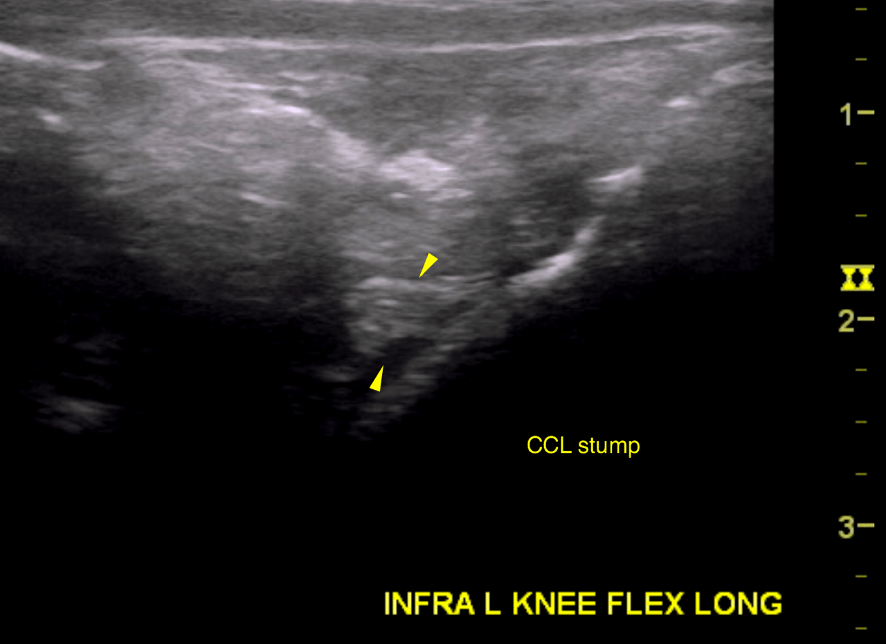
History of bilateral CCL injury, Had TTA on right knee Dec17/15, had bilateral U/S knees Dec 11/15. Pathology in left knee also noted. Played with puppy and now limping and favoring left stifle.
Physical Exam: Full Cr drawer sign- very loose left stifle, positive CTT, Now full rupture of CCL
History of bilateral CCL injury, Had TTA on right knee Dec17/15, had bilateral U/S knees Dec 11/15. Pathology in left knee also noted. Played with puppy and now limping and favoring left stifle.
Physical Exam: Full Cr drawer sign- very loose left stifle, positive CTT, Now full rupture of CCL