Ultrasound of both stifles-
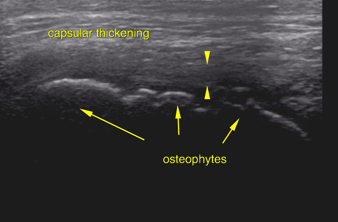
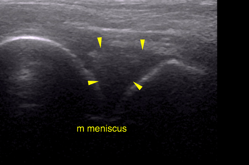
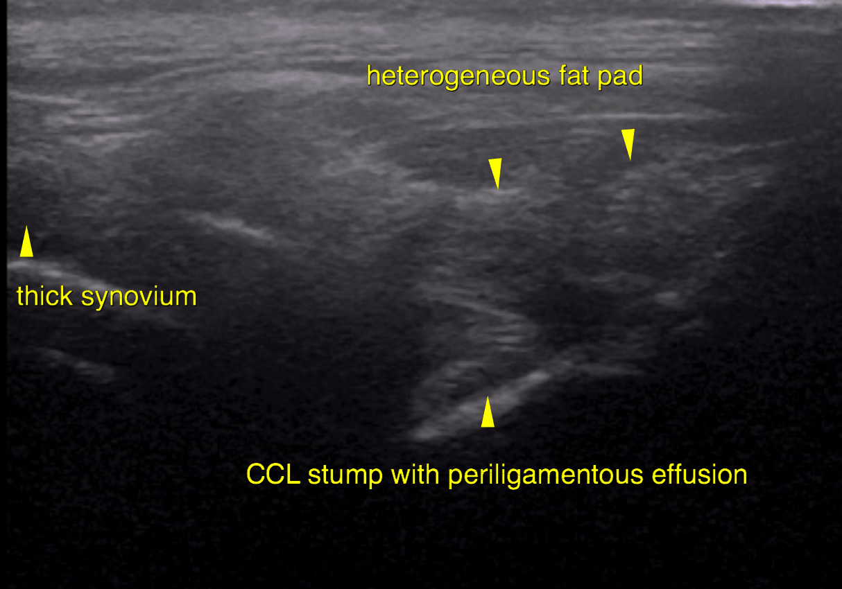
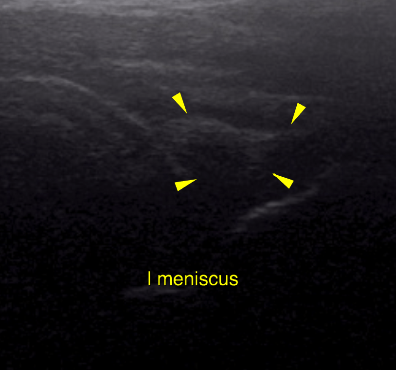
Left stifle: A moderate amount of anechoic effusion as well as moderate synovial proliferations and capsular thickening is noted within the suprapatellar and infrapatellar recess of the left stifle joint. There are moderate osteophyte formations at the periarticular margins of the stifle joint emphasizing the femoropatellar joint. Cartilage breakdown is noted at the femoral trochlea. The cranial cruciate ligament presents as a thick echogenic stump at the eminence of the tibia and is surrounded by a moderate amount of effusion and delineated by thick synovium. The visible portion of the medial meniscus is mildly heterogenous in echogenicity. There is no evidence of meniscal prolapse or rupture. The lateral meniscus is in situ with even surface and a moderate uniform overall increase in echogenicity.
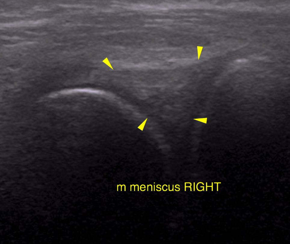
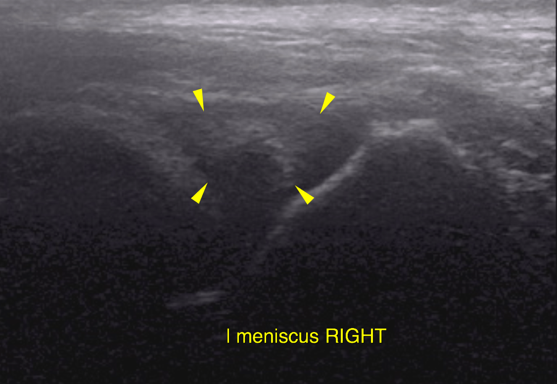
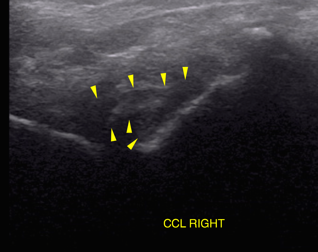
Right stifle: A mild amount of anechoic effusion as well as moderate synovial thickening is noted within the suprapatellar and infrapatellar recess of the right stifle joint. There are mild osteophyte formations at the periarticular margins of the femoropatellar and femerotibial joint. The cranial cruciate ligament is uneven in thickness, non-uniform in echogenicity an irregularly delineated. It appears to comprise a continuous and a discontinuous portion. The medial and lateral meniscuses are in situ with smooth outline and a mild uniform increase in overall echogenicity.