ORTHO – Degenerative joint disease (DJD) and full cranial cruciate ligament (CCL) rupture of the left stifle in a 8 year old MN Nova Scotia Duck Toller dog

Case Study

ORTHO – Degenerative joint disease (DJD) and full cranial cruciate ligament (CCL) rupture of the left stifle in a 8 year old MN Nova Scotia Duck Toller dog

This 8 year old MN Nova Scotia Duck Toller dog presented for right hind lameness of one month duration.

Physical exam – positive cranial drawer, possitive CCT, unstable joint

CBC/Chem WNL
This 8 year old MN Nova Scotia Duck Toller dog presented for right hind lameness of one month duration.

Physical exam – positive cranial drawer, possitive CCT, unstable joint

CBC/Chem WNL

Image Interpretation

Ultrasound of the right stifle – The supra- and infrapatellar recess of the right stifle joint contain a moderate and mild
amount of anechoic effusion respectively. The joint capsule reveals moderate
thickening. Moderate synovial proliferations are noted.
The cartilage layers as appreciated onto the femoral trochlea and the femoral condyles
are even, smooth, and anechoic as far as seen. Mild increased echogenicity is noted at
the periarticular zones only.
A moderate amount of osteophytes is noted at the periarticular margins of the
femoropatellar and a mild amount of osteophytes is noted at the femorotibial joint.
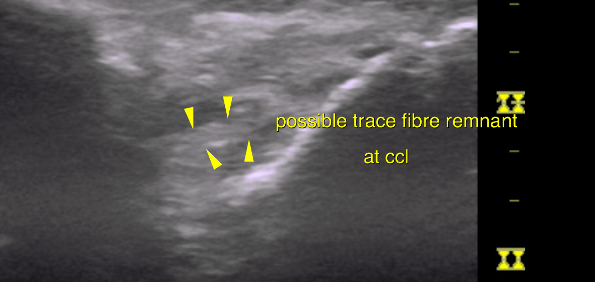
The cranial cruciate ligament (CCL) presents as an echogenic stump next to the distal
insertion at the intercondylar eminence of the tibia. There is scant periligamentous
effusion as well as a thick synovial fold and a mild amount of echogenic reactive
tissue. A possible fine trace of fibre remnants is seen in the region of the CCL but is
more consistent with reactive synovial tissue than actual remnants of the CLL.
The infrapatellar fat body presents heterogenous with multifocal increase in echogenicity as found commonly in chronic degenerative joint disease (DJD). 
The medial meniscus is well visible. It is in situ with even surface and uniform
echogenicity. There is no local effusion.
The lateral meniscus shows signs of mild degeneration. There is no evidence of
meniscal injury. The position is in situ. A mild to moderate amount of synovial
proliferation is noted within the lateral compartment of the femorotibial joint.

DX

The findings of the right stifle joint are compatible with moderate generalized chronic degenerative joint disease (DJD) with joint effusion, osteophytosis, capsular fibrosis and synovitis. Moreover the findings are consistent with rupture of the cranial cruciate ligament. Full rupture with complete biomechanical failure is assumed here.

Outcome

A medial meniscal injury is not seen. There are signs of lateral meniscal degeneration.

Patient Information

Patient Name : Hunter Southcott/Westview VH
Gender : Male, Neutered
Species : Canine
Type of Imaging : Ultrasound
Status : Complete
Liz Wuz Here : Yes

Clinical Signs

  • Lameness

Exam Finding

  • Doughy Abdomen

Images

bildschirmfoto_2016-02-12_um_12

Clinical Signs

  • Lameness