Ultrasound of the stifles –
Right:
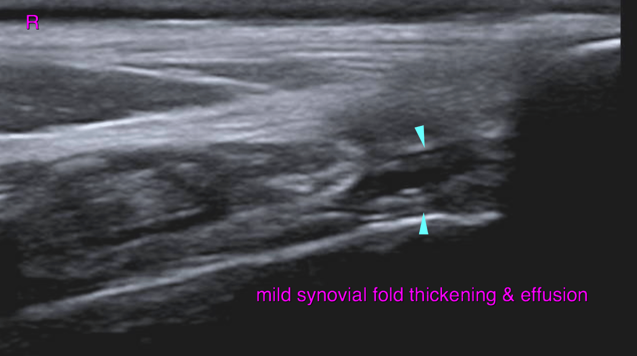
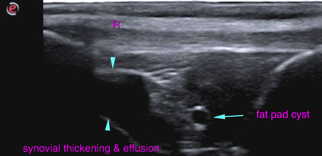
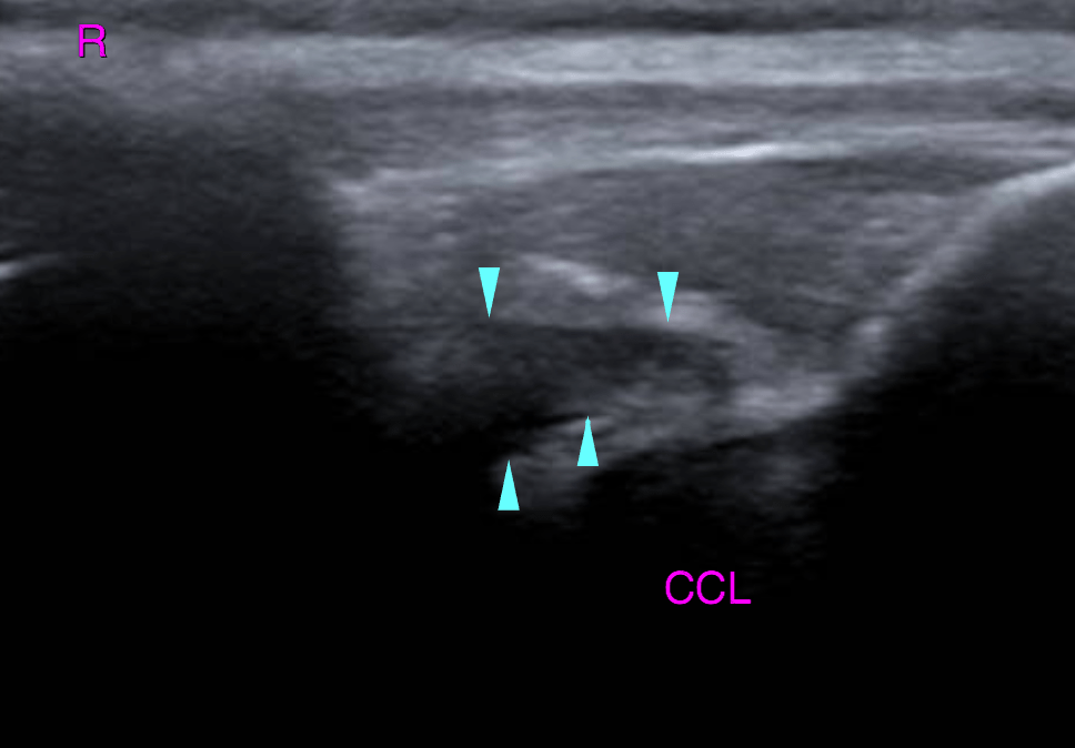
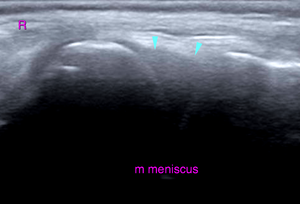
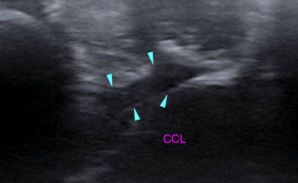
The suprapatellar recess presents mild thickening of the synovial lining with mild anechoic effusion. A moderate amount of anechoic effusion and moderate synovial thickening are noted within the infrapatellar recess of the stifle joint.Emerging periarticular osteophytes are noted at the medial aspect of the femorotibial joint. The infrapatellar fat pad presents mild heterogeneity with a solitary anechoic cyst. The cranial cruciate ligament (CCL) is continuous with even outline and regular echo pattern. Compared with the left side moderate generalized volume increase of the CCL is noted though
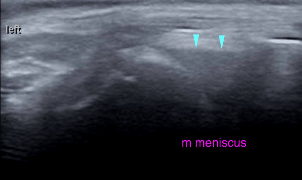
Left:
Emerging periarticular osteophytes are noted at the medial aspect of the femorotibial joint.