Skip to content
Customize Consent Preferences

We use cookies to help you navigate efficiently and perform certain functions. You will find detailed information about all cookies under each consent category below.

The cookies that are categorized as "Necessary" are stored on your browser as they are essential for enabling the basic functionalities of the site. ... 

Always Active

Necessary cookies are required to enable the basic features of this site, such as providing secure log-in or adjusting your consent preferences. These cookies do not store any personally identifiable data.

No cookies to display.

Functional cookies help perform certain functionalities like sharing the content of the website on social media platforms, collecting feedback, and other third-party features.

No cookies to display.

Analytical cookies are used to understand how visitors interact with the website. These cookies help provide information on metrics such as the number of visitors, bounce rate, traffic source, etc.

No cookies to display.

Performance cookies are used to understand and analyze the key performance indexes of the website which helps in delivering a better user experience for the visitors.

No cookies to display.

Advertisement cookies are used to provide visitors with customized advertisements based on the pages you visited previously and to analyze the effectiveness of the ad campaigns.

No cookies to display.

ORTHO – Cranial cruciate rupture with joint effusion, osteophytosis, capsular fibrosis, bone sclerosis in a 5 year old MN Labrador Retriever dog

Case Study

ORTHO – Cranial cruciate rupture with joint effusion, osteophytosis, capsular fibrosis, bone sclerosis in a 5 year old MN Labrador Retriever dog

The patient is a 5 year old MN Labrador Retriever dog presented for right hind lameness. TTA surgery right stifle in August of 2015. 

Physical Exam: positive cranial drawer, positive CTT, pain left stifle

CBC and Chem wnl

The patient is a 5 year old MN Labrador Retriever dog presented for right hind lameness. TTA surgery right stifle in August of 2015. 

Physical Exam: positive cranial drawer, positive CTT, pain left stifle

CBC and Chem wnl
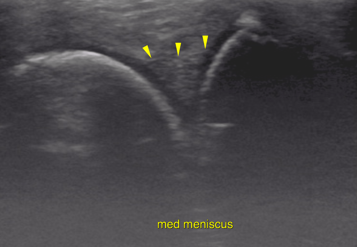
Image Interpretation

Left stifle joint:
The supra- and infrapatellar recesses and the tendon sheath of the long digital extensor
muscle (communicates with the joint) of the left stifle joint contain a moderate amount
of anechoic effusion. The joint capsule reveals moderate thickening. Moderate
synovial proliferations are noted.
The cartilage layers as appreciated within the patellar sulcus at the femoral trochlea
and onto the femoral condyles are widely irregular in outline and increased in
echogenicity. The subchondral bone of the femoral condyles reveals multifocal
irregularity.
A moderate amount of osteophytes is noted at the femoral trochlea next to the sulcus of
the patella and at the periarticular margins of the femorotibial joint.
An echogenic stump of disrupted fibers is seen at the distal insertion of the cranial
cruciate ligament (CCL). Moderate periligamentous effusion is present.
The infrapatellar fat body is heterogenous and increased in echogenicity. The medial meniscus is in situ with mildly undulating surface and uniform
echogenicity. No injury is seen. The lateral meniscus is in situ with even surface and
uniform echogenicity. No meniscal disruption is seen. A moderate amount of
echogenic fibrous tissue is seen superficial/lateral to the lateral meniscal surface.

DX

The left stifle joint is affected by moderate generalized chronic degenerative joint disease with joint effusion, cartilage breakdown, subchondral bone sclerosis, osteophytosis, synovial proliferation and capsular fibrosis. The ultrasonographic findings are compatible with cranial cruciate ligament pathology with biomechanical failure due to complete rupture. A meniscal tear is not seen.

Video

Patient Information

Patient Name : Roger Jukes
Gender : Male, Neutered
Species : Canine
Type of Imaging : Ultrasound
Status : Complete
Liz Wuz Here : Yes

Clinical Signs

  • Lameness

History

  • Cruciate ligament injury

Exam Finding

  • Cranial drawer posiive
  • Pain

Images

bildschirmfoto_2015-12-16_um_21bildschirmfoto_2015-12-16_um_22bildschirmfoto_2015-12-16_um_22bildschirmfoto_2015-12-16_um_22

Clinical Signs

  • Lameness