This 6 year old MN Belgian Shepherd dog has a history of intermittent lameness of over 1 year duration. Worsens with exercise. Decreased muscle mass right hind. Positive cranial drawer sign and CTT, moderate medial buttress sign.
Evaluate right stifle prior to TTA stabilization
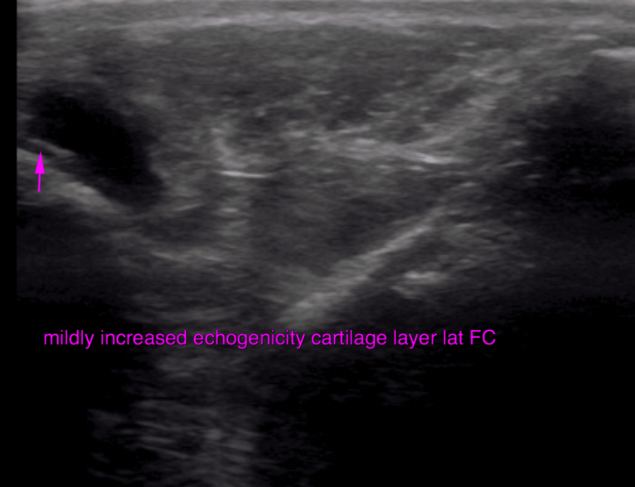
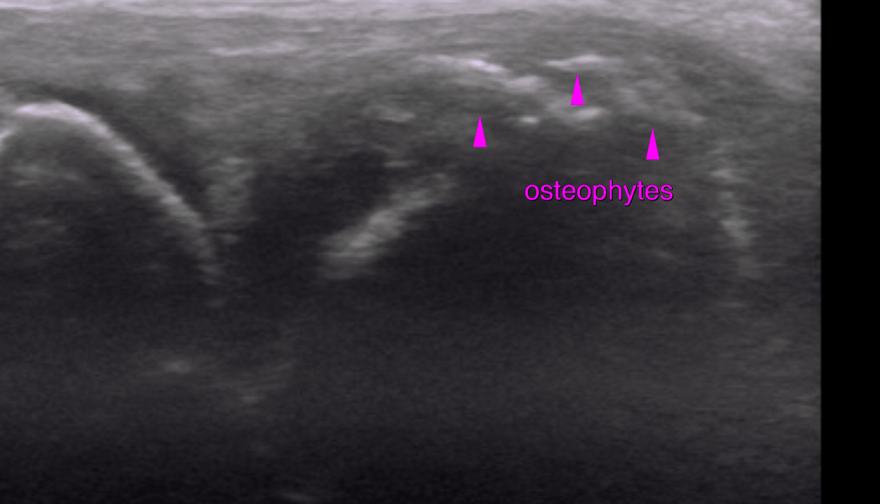
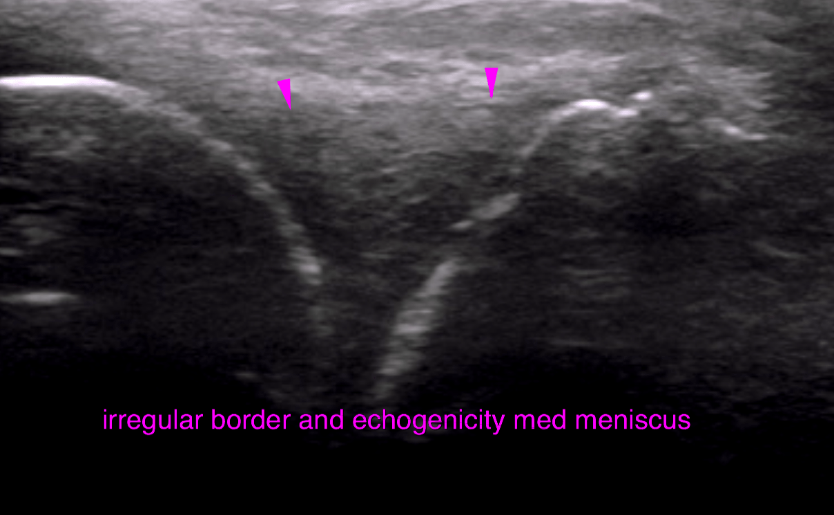
This 6 year old MN Belgian Shepherd dog has a history of intermittent lameness of over 1 year duration. Worsens with exercise. Decreased muscle mass right hind. Positive cranial drawer sign and CTT, moderate medial buttress sign.
Evaluate right stifle prior to TTA stabilization