Mural Gastric Abscess with extensive Pancreatitis in a 12 year old FS Schnauzer

Case Study

Mural Gastric Abscess with extensive Pancreatitis in a 12 year old FS Schnauzer

A 12-year-old SF Schnauzer that had been treated for pancreatitis was presented for ongoing inappetence and vomiting. Abnormalities on serum biochemistry were azotemia and elevated amylase and lipase. Survey abdominal radiographs showed decreased abdominal detail.

A 12-year-old SF Schnauzer that had been treated for pancreatitis was presented for ongoing inappetence and vomiting. Abnormalities on serum biochemistry were azotemia and elevated amylase and lipase. Survey abdominal radiographs showed decreased abdominal detail.

DX

Mural gastric abscess with extensive pancreatitis

Sonographic Differential Diagnosis

Mural gastric abscess.
Extensive pancreatitis was noted.
Renal and bladder calculi.

Image Interpretation

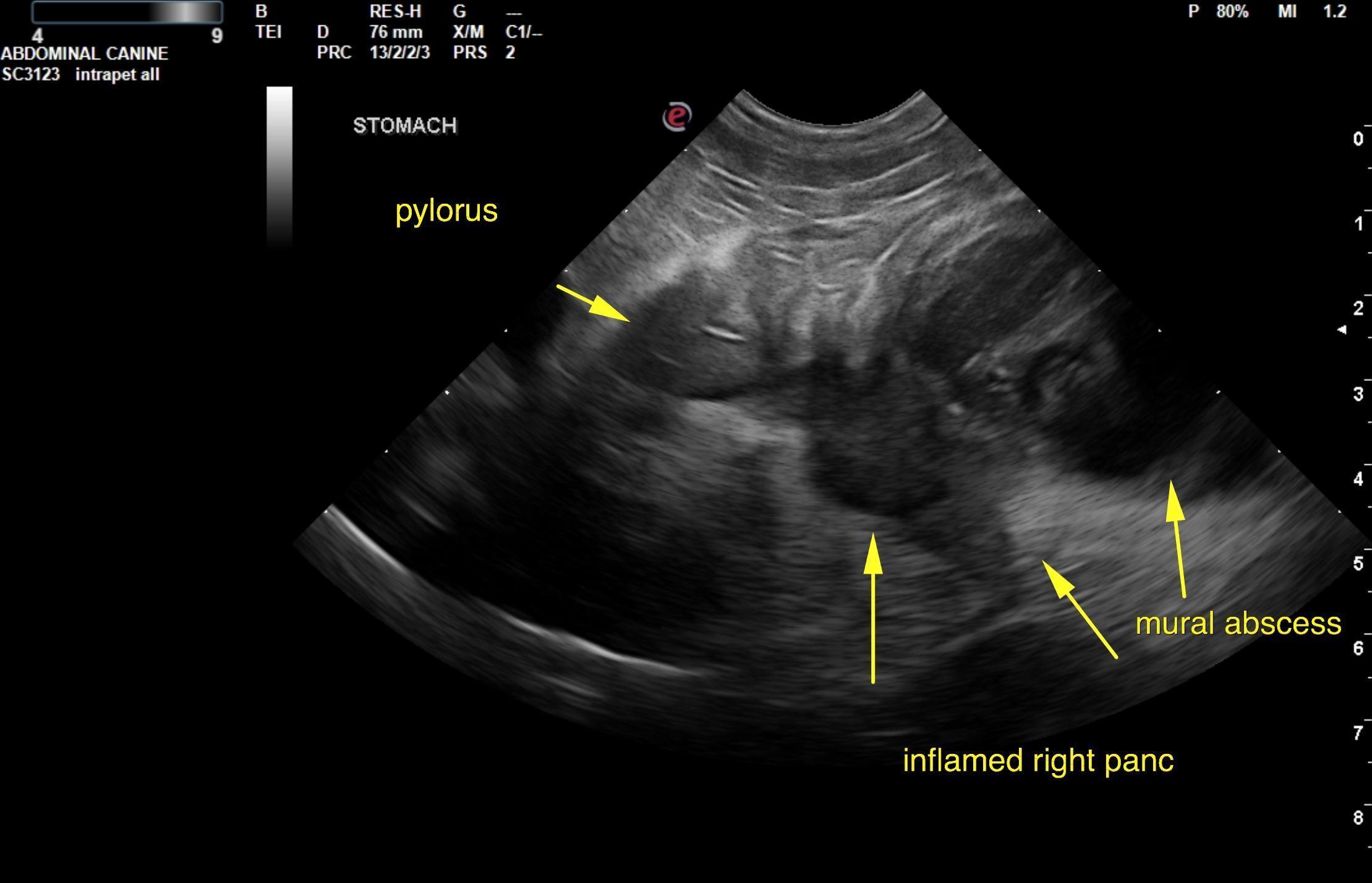
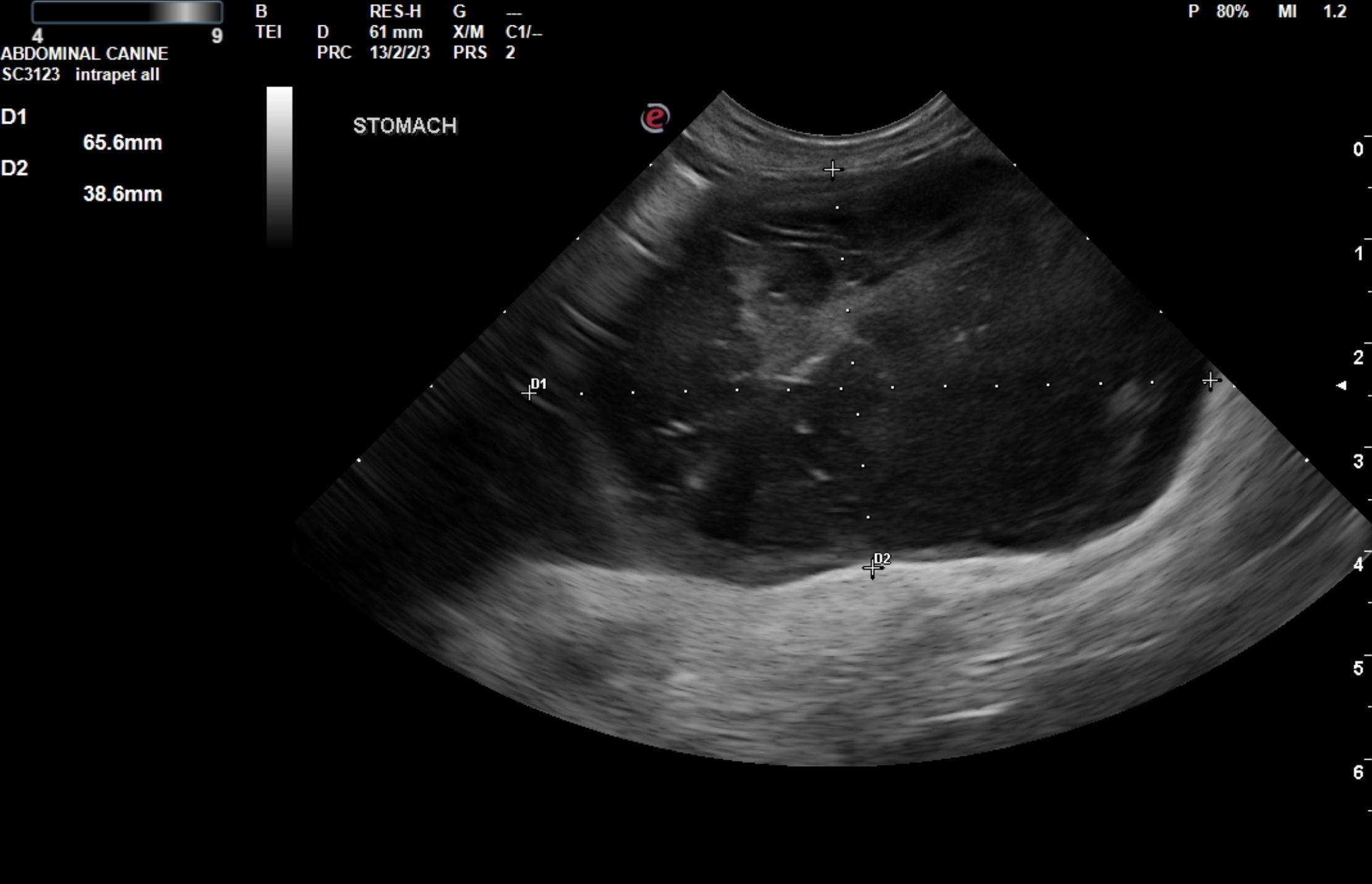
The stomach in this patient presented a 6.5 x 3.8 cm mural abscess.  The abscess appeared to be in the greater curvature. The pylorus was thickened.

The right limb of the pancreas was hypoechoic with hyperechoic surrounding fat.

The urinary bladder wall itself was unremarkable. Multiple, non obstructive bladder calculi were noted and measured 0.5 cm and 0.6 cm. No evidence of inflammatory or neoplastic changes were noted. The ureters were not visible and therefore considered normal.

The kidneys revealed largely normal size and structure, corticomedullary definition and ratio (cortex 1/3 of medulla) were essentially maintained with some age related loss of curvilinear patterns. The cortices presented largely uniform texture with some age related echogenic changes that are not likely of clinical significance at this time unless inflammatory sediment or proteinuria is present. Medullary echogenicity differed distinctly from that of the cortex and no evidence or dilation could be seen. The capsules were acceptably uniform for this age patient without dramatic irregularities. Renal calculi were noted.  The right kidney measured 5.1 cm. The left kidney measured 4.81 cm.
Outcome

Immediate exploratory surgery with expectations of resecting a portion of the gastric wall with abdominal lavage, treating for peritonitis, cystotomy, stone analysis and culture, was recommended. Expression of the gallbladder was recommended at the time of surgery. Underlying neoplasia is not obvious, yet could not be completely ruled out. Minor, epigastric lymphadenopathy was noted 1.0 cm. Surgical biopsies of the stomach would be recommended. Very guarded prognosis. A recheck sonogram was recommended in 3-5 days to ensure adequate resolution.

Clinical Differential Diagnosis

Renal – acute kidney injury, renolith, abscess, pyelonephritis
Pancreas – pancreatitis, neoplasia, abscessation
GIT – foreign body, neoplasia, ulceration, focal perforation with peritonitis

Patient Information

Patient Name : Ginger Sheets
Gender : Female, Spayed
Species : Canine
Type of Imaging : Ultrasound
Status : Complete
Liz Wuz Here : Yes
Code : 04_00558

Clinical Signs

  • Anorexia
  • Vomiting

History

  • Pancreatitis

Images

rb1ap6

Blood Chemistry

  • Amylase, High
  • Azotemia
  • Lipase, High

Clinical Signs

  • Anorexia
  • Vomiting
Skip to content