Left adrenal mass and left liver mass in a 12 year old MN DLH cat

Case Study

Left adrenal mass and left liver mass in a 12 year old MN DLH cat

A 12-year-old NM DLH was presented for evaluation of one-month duration of hiding in strange places, inappropriate urination, and PuPD. On physical examination, an abdominal mass near the cranial pole of the left kidney and grade 2/6 left parasternal systolic heart murmur were present. Abnormalities on CBC and serum biochemistry were leukocytosis, FIV positive, hypokalemia (3.6) and azotemia (BUN 48, creatinine 2.6).
A 12-year-old NM DLH was presented for evaluation of one-month duration of hiding in strange places, inappropriate urination, and PuPD. On physical examination, an abdominal mass near the cranial pole of the left kidney and grade 2/6 left parasternal systolic heart murmur were present. Abnormalities on CBC and serum biochemistry were leukocytosis, FIV positive, hypokalemia (3.6) and azotemia (BUN 48, creatinine 2.6).
DX

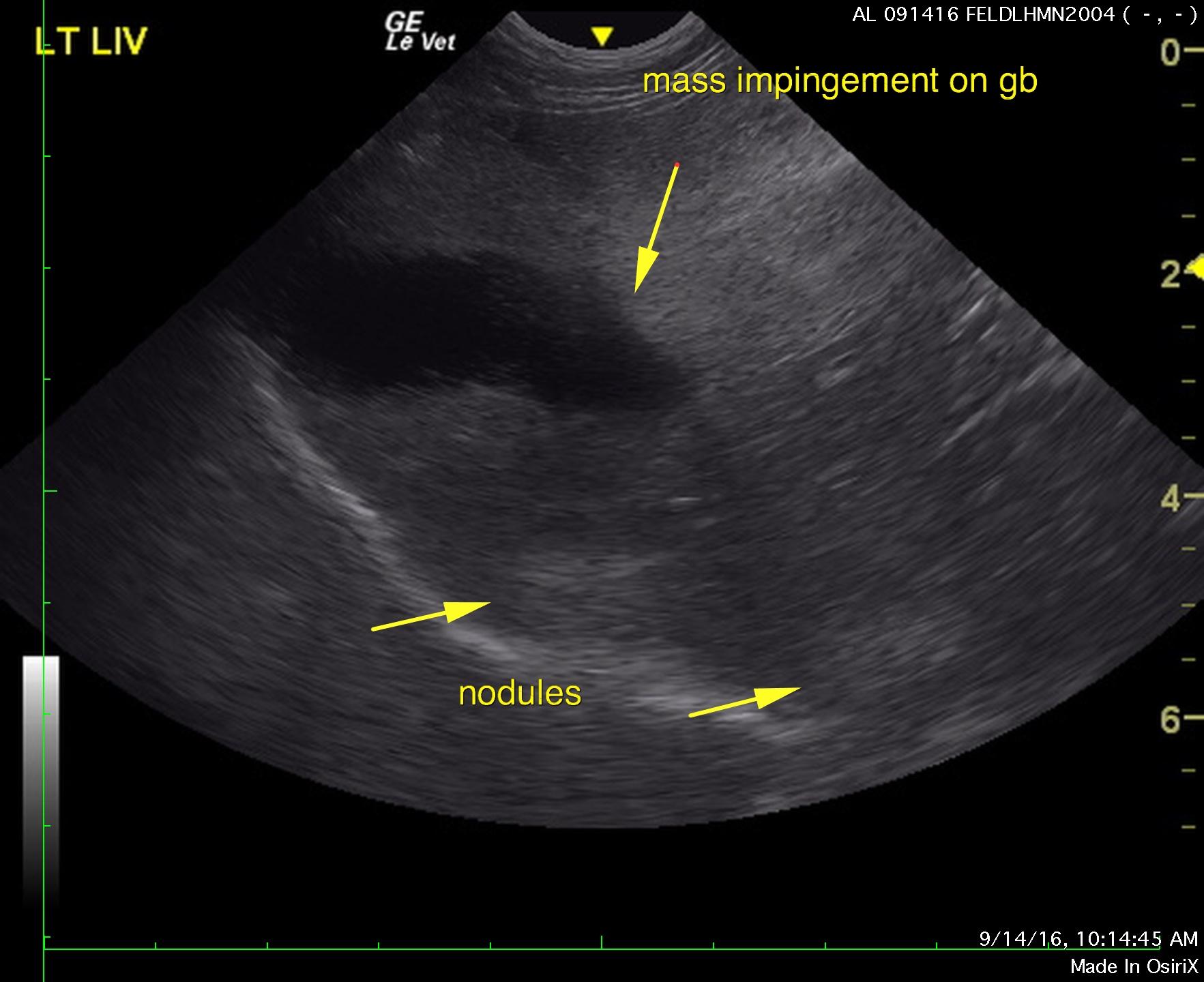
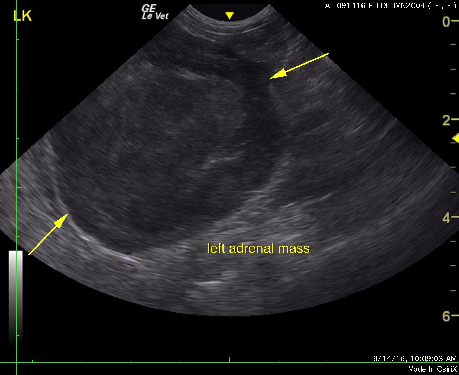
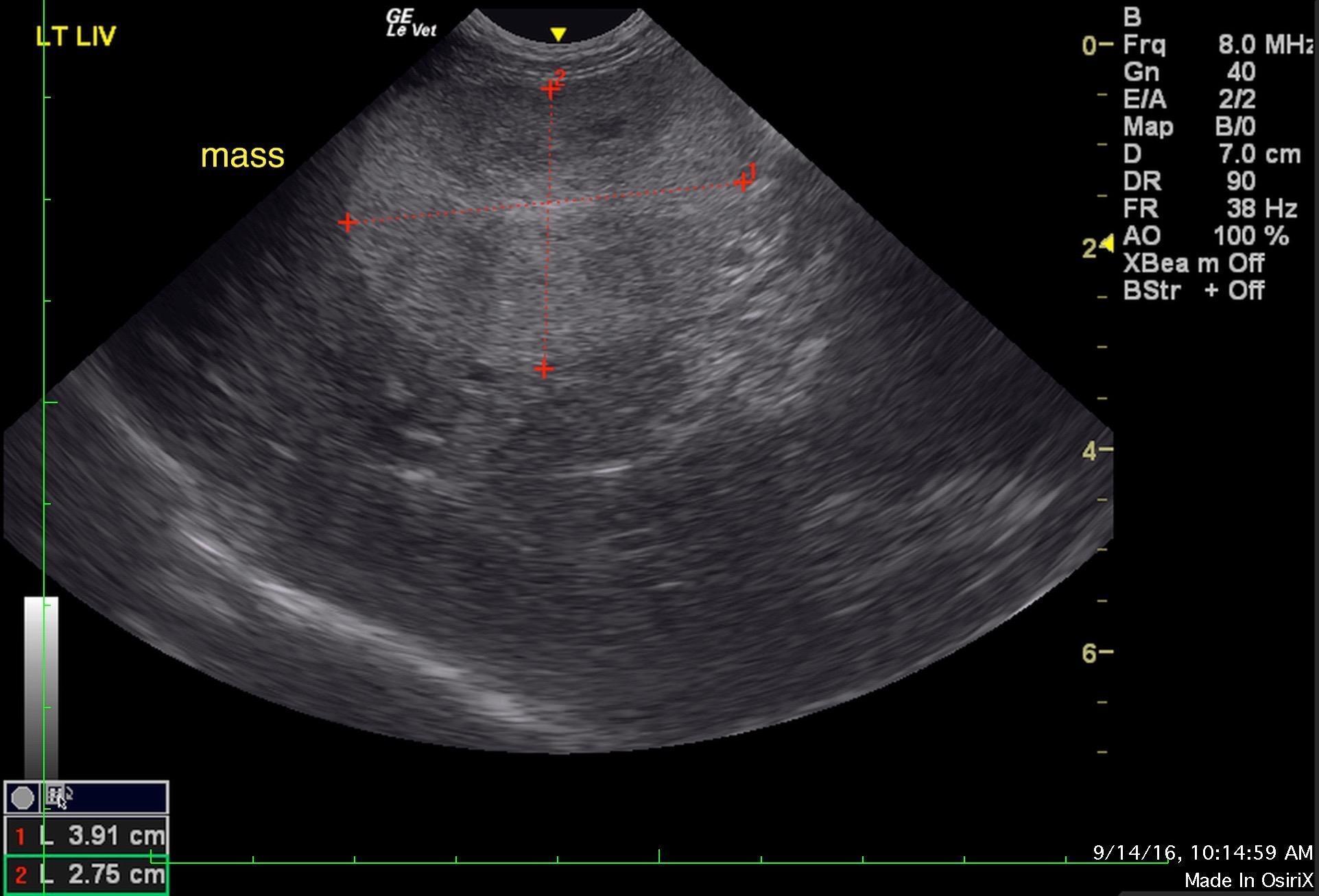
Mass in the region of the left adrenal gland. Left-sided liver mass. Undefined occasional hepatic nodules, appear benign.

Sonographic Differential Diagnosis

Mass in the region of the left adrenal gland.
Left-sided liver mass. Undefined occasional hepatic nodules, appear benign.

Image Interpretation

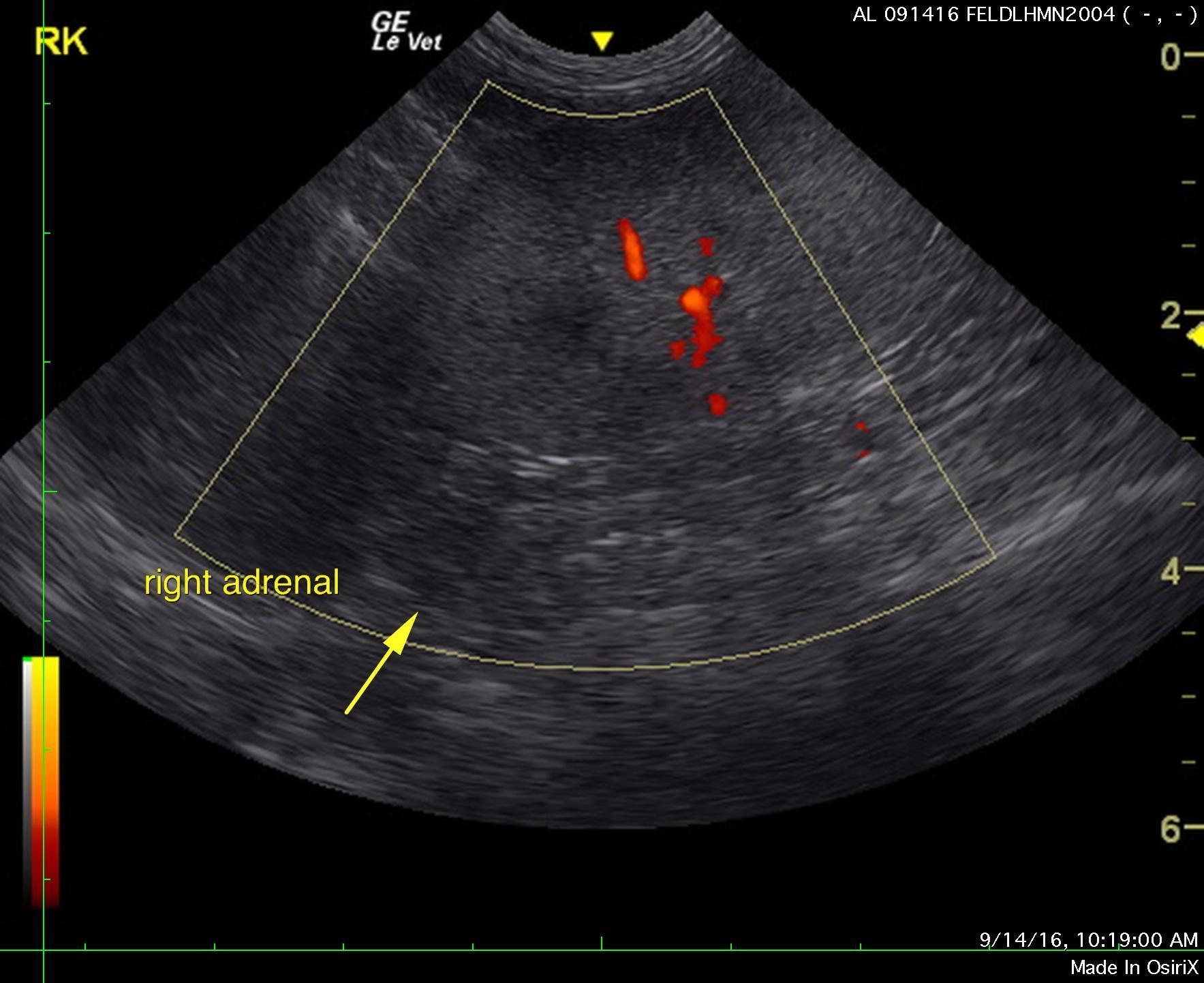
A left adrenal gland mass was noted in this patient and measured 4.0 cm. This impinged upon the left kidney. A trace amount of free fluid was noted. Adhesion to the left kidney is likely an issue. Surrounding vascularity appeared free of invasion. This appears resectable. The right adrenal gland was visualized and appeared normal. 

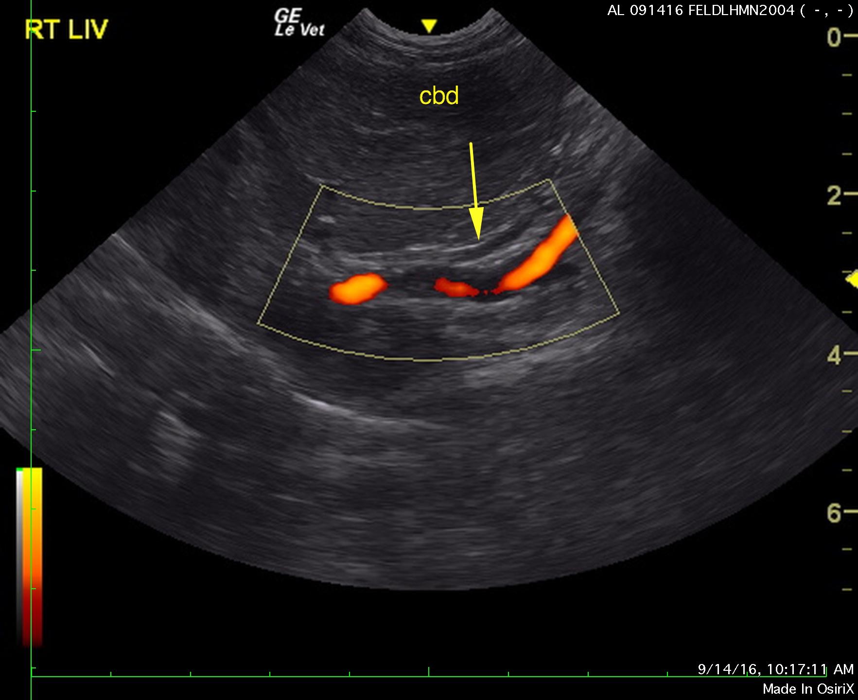
The left liver revealed a hyperechoic mass that measured 3.91 x 2.75 cm. The liver mass appears isolated and encapsulated in the region of the left adrenal gland. The mass impinges upon the gallbladder. Other hyperechoic nodules were noted in the right liver. These appear subjectively benign. The remainder of the liver revealed mildly increased portal markings and remodeling. The gallbladder was unremarkable.  The common bile duct was normal as was the portal vein and vena cava. 

Outcome

CT evaluation would be ideal in this patient for surgical planning. Aldosterone level is recommended in this patient to assess for Conn’s syndrome if this is an adrenal mass. It is occupying the region of the adrenal and the renal displacement would suggest adrenal mass. Ultrasound-guided FNA could be considered; however, direct surgical exploratory with expectations towards left liver lobectomy and adrenalectomy would be recommended. Subjectively the left adrenal mass appears to be relatively benign. Low grade hepatocellular carcinoma is suspected or cystadenoma. The mass in the region of the left adrenal gland appears to be relatively aggressive, yet encapsulated. There was no obvious invasion into the adjacent vena cava noted. Serial blood pressure measurements and ECG would be warranted given the arrhythmogenic activity.

Clinical Differential Diagnosis

Renal – renal neoplasia, granuloma, abscess, hematoma, chronic likely disease, pyelonephritis
Adrenal neoplasia – hyperaldosteronism
Cardiac – cardiomyopathy

Patient Information

Patient Name : Herman Bartali
Gender : Male, Neutered
Species : Feline
Type of Imaging : Ultrasound
Status : Complete
Liz Wuz Here : Yes
Code : 07_00194

Clinical Signs

  • Hiding behavior
  • Inappropriate Urination
  • PU-PD

Exam Finding

  • Heart Murmur
  • Palpable mass

Images

as4as9as3_1as7as8

Blood Chemistry

  • Azotemia
  • Potassium, Low

CBC

  • WBC, High

Clinical Signs

  • Hiding behavior
  • Inappropriate Urination
  • PU-PD

Special Testing

  • FIV Positive
Skip to content