A 10-year-old FS DSH cat was presented for weight loss and a possible abdominal mass. Abnormalities on blood chemistry were hypoalbuminemia and elevated ALP.
A 10-year-old FS DSH cat was presented for weight loss and a possible abdominal mass. Abnormalities on blood chemistry were hypoalbuminemia and elevated ALP.
Case Study
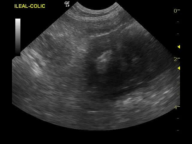
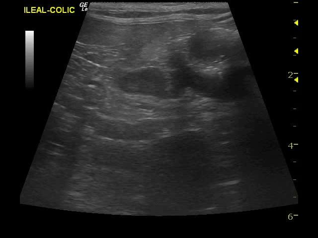
Intestinal lymphoma in ileocecal mass in a 10-year-old FS DSH cat
Sonographic Differential Diagnosis
Ileocecocolic mass with possible splenic involvement. Likely lymphoma or adenocarcinoma, leiomyosarcoma, and minor possibility of complicated inflammatory bowel disease.
Image Interpretation
The gastrointestinal tract revealed a mural ileocecocolic mass with deviation of the lumen. The mass measured 2 x 3 cm. This mass by itself appears resectable. It was localized in the ileocecocolic area. However, associated lymphadenopathy that measured 1.6 x 0.7 cm was also noted as well as other small lymph nodes in the area.
DX
Large cell, lymphoblastic lymphosarcoma.
Outcome
The owners were considering oncology referral vs. euthanasia at last communication.
Comments
The mass appears resectable.Fine-needle aspirates of the spleen and mass would be warranted for a definitive diagnosis,
Clinical Differential Diagnosis
Multiple organ pathology -GI tract/spleen/liver/renal (neoplasia, granuloma, abscess). Splenic pathology (infarction/torsion). Renal pathology (hydronephrosis)
Sampling
Microscopic Interpretation: LYMPHOSARCOMA, LARGE CELL/LYMPHOBLASTIC FORM Comment: This is a diagnosis that warrants a cautious prognosis. Large cell lymphosarcoma that involves the G.I. tract typically responds poorly to chemotherapy while cases that do not have G.I. involvement will often respond with 6-18 month disease free intervals with chemotherapy.It is suspected that the ileocecal lymph node was aspirated, and if the tumor is confined to the node only chemotherapy will likely prove beneficial. Unfortunately there is a high probability of concurrent G.I. involvement as this is a common region for G.I. lymphosarcoma to originate.
Video
Patient Information
Patient Name :
Gunny S
Gender :
Female, Spayed
Species :
Feline
Type of Imaging : Ultrasound
Status :
Complete
Liz Wuz Here :
Yes
Code :
04_00255
Clinical Signs
- Palpable Mass
- Weight loss
History
- Weight Loss
Exam Finding
- Palpable mass
Images
Blood Chemistry
- Albumin, Low
- Alkaline Phosphatase (SAP), High
Clinical Signs
- Palpable Mass
- Weight loss