This 12-year-old FS DSH cat presented for weight loss. The physical examination revealed a palpable abdominal mass and poor body condition. A CBC and blood chemistry profile revealed only a moderate neutrophilia with a left shift.
This 12-year-old FS DSH cat presented for weight loss. The physical examination revealed a palpable abdominal mass and poor body condition. A CBC and blood chemistry profile revealed only a moderate neutrophilia with a left shift.
Case Study
Intestinal carcinoma in a 12 year old FS DSH cat
Sonographic Differential Diagnosis
The differential diagnoses included an obstructing annular bowel wall mass with suspect adjacent mesentery involvement. These findings were considered consistent with neoplasia, most notably adenocarcinoma.
Image Interpretation
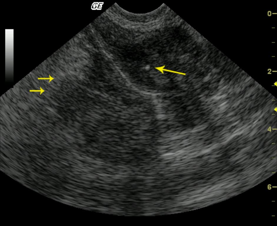
A segment of small bowel has a markedly, circumferentially thickened wall (image 1). The peripheral margins of the intestinal mass are multilobulated, with subtle finger-like projections surrounded by mildly echogenic omentum and mesentery, suggesting serosal layer interruption (short arrows.) The ovoid, echogenic focus visualized within the mass at the 1 o`clock position represents the beveled surface of a 22-gauge needle (long arrow.) Continuous hypoechoic mass is visible within the bowel wall indicated by the hyperechoic luminal interface indicating that this mass derives from the intestine. Mural detail is completely lost (Video 1.)
DX
Intestinal carcinoma
Outcome
The patient was euthanized due to poor therapeutic response and prognosis.
Clinical Differential Diagnosis
Neoplasia, aggressive inflammatory GI disease with lymphadenopathy, pancreatitis with mass lesion, foreign body.
Sampling
A 22-ga US-guided FNA (Video 2) was obtained, and cytology was consistent with intestinal carcinoma with suppurative inflammation.
Video
Patient Information
Patient Name :
Jill P
Gender :
Female, Spayed
Species :
Feline
Type of Imaging : Ultrasound
Status :
Complete
Liz Wuz Here :
Yes
Code :
04_00018
Clinical Signs
- Weight loss
History
- Weight Loss
Exam Finding
- Palpable mass
- Weight loss
Images
CBC
- Left Shift
- WBC, High
Clinical Signs
- Weight loss