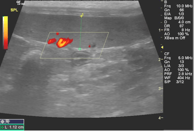
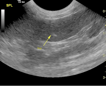
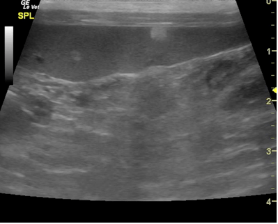
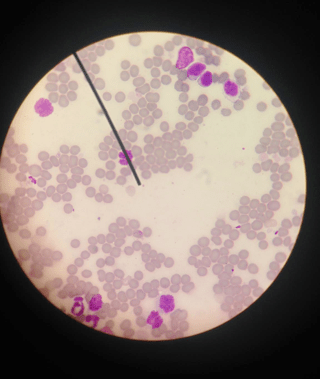
A 16-year-old SF DSH cat was presented for evaluation. Abnormalities on serum biochemistry were azotemia (BUN 43, creatinine 3.2) and elevated liver enzyme activity (ALT 443, ALP 151). Survey radiographs revealed hyper inflated lungs and excessive upper gastrointestinal gas.
A 16-year-old SF DSH cat was presented for evaluation. Abnormalities on serum biochemistry were azotemia (BUN 43, creatinine 3.2) and elevated liver enzyme activity (ALT 443, ALP 151). Survey radiographs revealed hyper inflated lungs and excessive upper gastrointestinal gas.