Inflamed gallbladder mucocele in an 8 year old FS Miniature Schnauzer

Case Study

Inflamed gallbladder mucocele in an 8 year old FS Miniature Schnauzer

An 8-year-old FS Miniature Schnauzer was presented for evaluation of elevated liver enzyme activity on routine pre-anesthetic blood work. Physical examination was normal. Abnormalities on CBC and serum biochemistry were mild non-regenerative anemia (37%) and elevated ALT (295) and pre-and postprandial bile acids 101 and 98, respectively. cPL was within reference range.
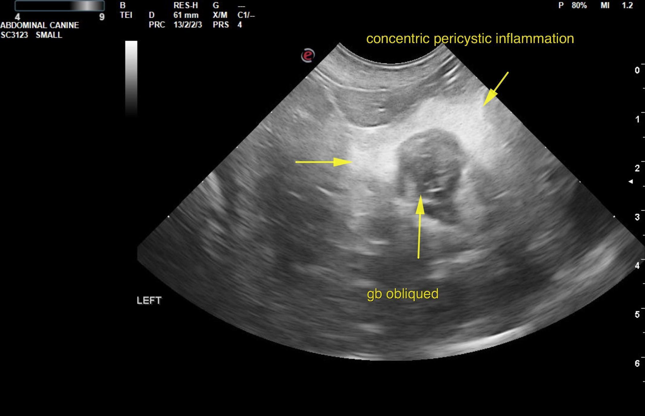
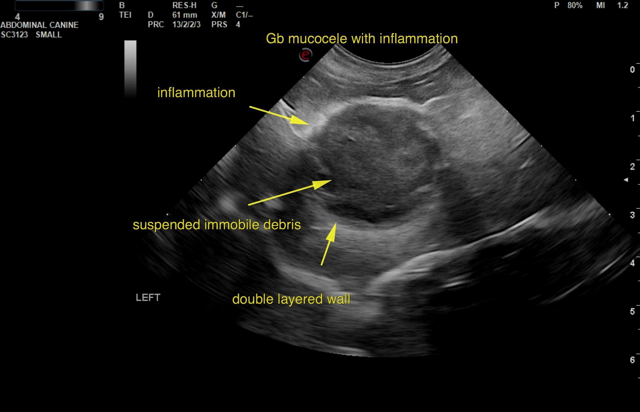
An 8-year-old FS Miniature Schnauzer was presented for evaluation of elevated liver enzyme activity on routine pre-anesthetic blood work. Physical examination was normal. Abnormalities on CBC and serum biochemistry were mild non-regenerative anemia (37%) and elevated ALT (295) and pre-and postprandial bile acids 101 and 98, respectively. cPL was within reference range.

DX

Inflamed gallbladder mucocele

Sonographic Differential Diagnosis

Inflamed gallbladder mucocele.
Mild bilateral pyelectasia.

Image Interpretation

The liver was mildly enlarged with uniform vacuolar hepatopathy pattern. However, the gallbladder in this patient presented an inflamed gallbladder mucocele. A double layered wall and suspended immobile debris were noted. Concentric inflammation was noted completely around the gallbladder. Minor, heterogenous parenchymal changes were noted. This is a surgical emergency. The common bile duct did not appear to be involved.

Outcome

This is a surgical emergency. Immediate cholecystectomy is recommended. .Cholecystectomy was performed in this patient .

Clinical Differential Diagnosis

Gall bladder – mucocele, cholecystitis, neoplasia
Liver – chronic-active hepatitis, neoplasia, reactive hepatopathy, nodular regeneration, cirrhosis

Sampling

Liver and gallbladder biopsy was performed. The gallbladder biopsy revealed a mucocele with chronic ulcerative/necrotizing, mixed, cellular and fibrosing transmural cholecystitis. Liver biopsies revealed mild, multifocal lymphoplasmacytic portal hepatitis and diffuse, moderate, hepatocellular vacuolar degeneration.

Patient Information

Patient Name : Sophia Kilcup, Ruidoso AC
Gender : Female, Spayed
Species : Canine
Type of Imaging : Ultrasound
Liz Wuz Here : Yes

Clinical Signs

  • Anemia

History

  • Elevated ALT
  • Elevated Bile Acids

Images

rui3rui4

Blood Chemistry

  • ALT (SGPT), High
  • Post-Prandial Bile Acids, High
  • Pre-Prandial Bile Acids, High

Clinical Signs

  • Anemia
Skip to content