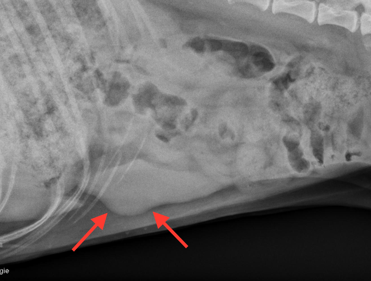
It is likely that the radiographic appearance was created by a positional artifact highlighting one of the mild surface protrusions.
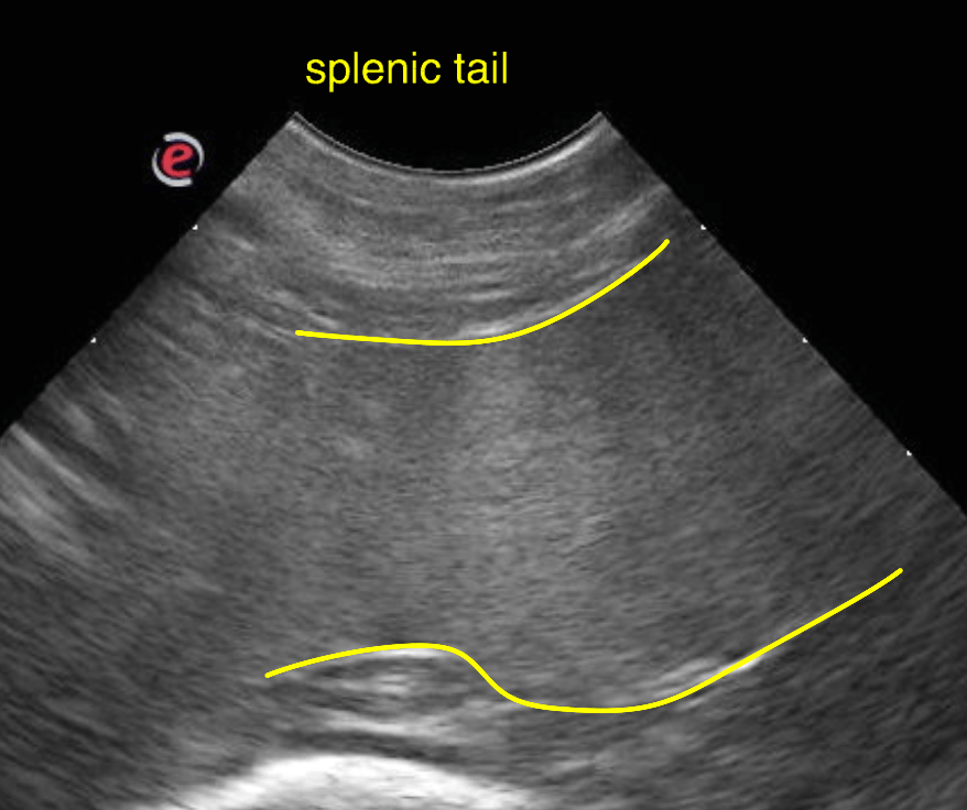
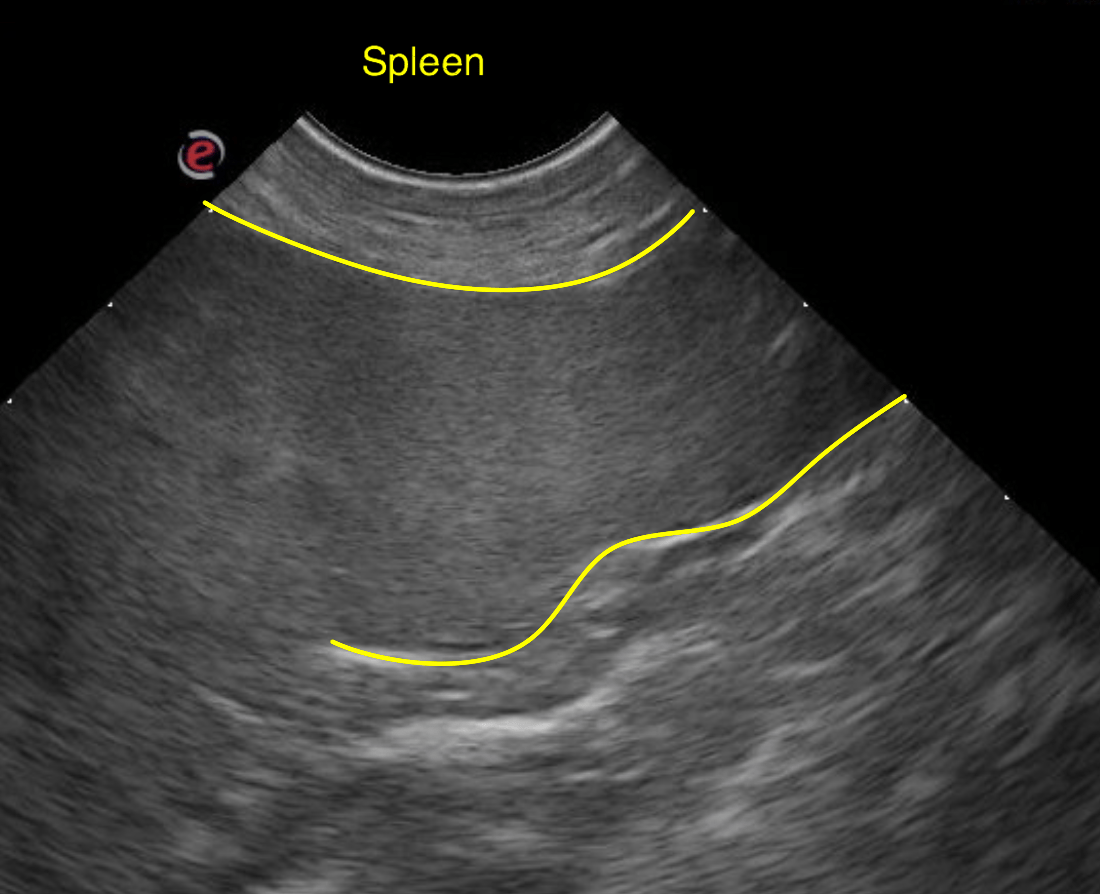
Yet disappearance of splenic nodules/masses is known to occur for benign lesions – such as foci of extramedullary hematopoiesis, nodular hyperplasia, hematoma, hemangioma – as well as highly vascularized malignancies – such as hemangiosarcoma – which can disappear when the spleen is volume contracted or during marked systemic hypotension.
Volume contraction of the spleen can be ruled out here based on the relatively large volume of the spleen. So if the patient was not severely hypotensive during the examination a nodular/mass like splenic malignancy is highly unlikely in this case.
A recheck ultrasonographic examination within the next 10 – 14 days should be considered to re-confirm the absence of transient splenic masses. At this time ultrasound guided fine needle aspiration may be performed to rule out a round cell neoplastic infiltrate as this is another possible reason for generalized hypersplenism even though the potential is low.