A 16-year-old MN DSH cat with a history of weight loss and previous inflammatory bowel disease was presented for poor appetite. Abnormalities on physical examination were dehydration, dental disease, and nasal discharge. Hematuria was present on urinalysis. Initial CBC showed leukocytosis and anemia, which remained fairly unchanged after one month of antibiotic therapy.
A 16-year-old MN DSH cat with a history of weight loss and previous inflammatory bowel disease was presented for poor appetite. Abnormalities on physical examination were dehydration, dental disease, and nasal discharge. Hematuria was present on urinalysis. Initial CBC showed leukocytosis and anemia, which remained fairly unchanged after one month of antibiotic therapy.
Case Study
IBD, TCC, splenic mastocytosis in a 16 year old MN DSH cat
Sonographic Differential Diagnosis
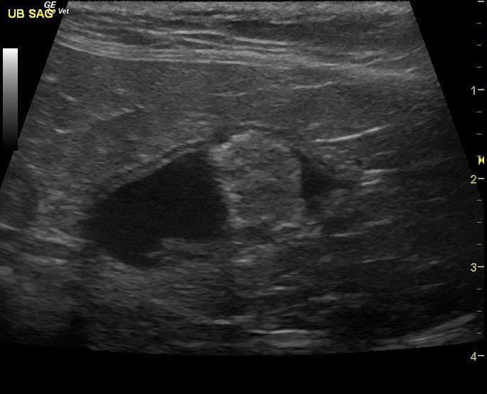
Probable benign dysfunctional bowel. Bladder pathology (transitional cell carcinoma or other neoplastic). Foreign body. Low grade hepatopathy.
Image Interpretation
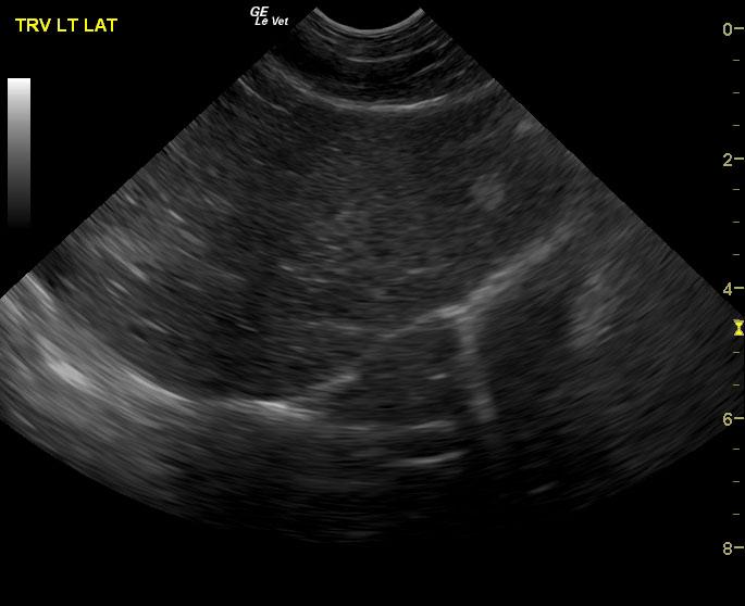
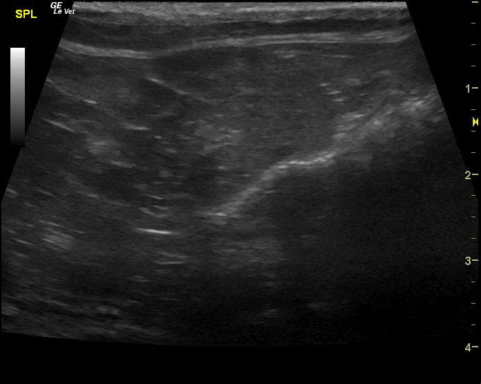
The gastrointestinal tract was largely unremarkable. However, there were areas of dilated small intestine or echogenic debris and gas artifact. Areas of empty small intestine were noted. Slight reactive omentum. Colonic artifact was also noted. This is indicative of adequate transit. Echogenic changes are noted associated with the wall and periserosal region. No overt foreign body was noted, yet could not be ruled out based on images provided due to gas artifact occluding any potential foreign matter.The bladder presented a 1.38 cm strongly mineralizing mass in the dorsal apex. This appears resectable.. The liver in this patient was largely normal in structure. The gallbladder and common bile duct were unremarkable. Slight areas of increased portal markings were noted with a hyperechoic nodule in the left lateral lobe which measured 0.6 cm with loss of structural architecture. The contour was mildly swollen. Nodular splenic changes.
DX
IBD, TCC, splenic mastocytosis
Outcome
Cat doing ok for now.
Comments
Full intestinal palpation would be recommended to assess the possibility of a foreign body in this region. However, if the bladder mass is to be resected, then exploratory of the jejunum where echogenic fat and fluid dilatation are noted could be performed to assess the possibility of a portion of dysfunctional bowel with potential transmural pathology that may need to be resected.
Clinical Differential Diagnosis
weight loss, anorexia – neoplasia, ulceration, foreign body, IBD, abscessation/granulomatous disease, pancreatic pathology ( pancreatitis, neoplasia). Hematuria- renal pathology ( interstitial cystitis, neoplasia, uroliths, pyelonephritis, renoliths) Age-related dental disease
Sampling
None
Video
Patient Information
Patient Name :
Tiger H
Gender :
Male, Neutered
Species :
Feline
Type of Imaging : Ultrasound
Status :
Complete
Liz Wuz Here :
Yes
Code :
04_00281
Clinical Signs
- Anorexia
- Dehydration
- Weight loss
History
- IBD
Exam Finding
- Dehydration
Images
CBC
- RBC, Low
- WBC, High
Clinical Signs
- Anorexia
- Dehydration
- Weight loss
Urinalysi
- Blood Present