Hyperadrenocorticism, phrenic thrombosis, and adrenal necrosis in a 10 year old FS mixed breed dog

Case Study

Hyperadrenocorticism, phrenic thrombosis, and adrenal necrosis in a 10 year old FS mixed breed dog


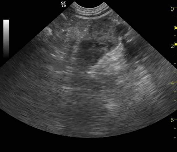
12/2005: A 10 year old FS mixed breed dog with a 2 year history of pituitary dependent HAC, medically well controlled with mitotane, was presented for a routine recheck exam and sonogram. Her blood chemistry revealed hyperglobulinemia, hyperphosphatemia, hypertriglyceridemia, and hyperamylasemia. The CBC was within normal limits. 

6/2006: The patient returned for her annual examination. Her blood chemistry revealed an elevated ALT enzyme activity, hyperphosphatemia, hyperamylasemia, and hypertriglyceridemia. The CBC was within normal limits. A urinalysis showed a high pH, but the urine microalbumin was high. The urine culture was negative. A T4 and free T4 were both within the normal reference range. An adrenal panel performed by the University of Tennessee found elevated baseline cortisol and elevated androstenedione concentrations. There was elevated post ACTH cortisol, elevated progesterone, elevated 17-OHP, and elevated aldosterone, thereby indicating increased adrenal activity. 

7/2006: An ACTH stimulation test was performed one month later, and results were consistent with hypoadrenocorticsm. She was treated with fludrocortisone and steroids.

8/2006: An ACTH stimulation test was performed one month later. The results were consistent with a diagnosis of iatrogenic hypoadrenocorticism.

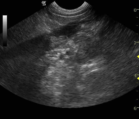
11/2006: A blood chemistry performed a few months later revealed hyperphosphatemia, hypertriglyceridemia, hyperlipasemia, and hyperamylasemia. The CBC and TT4 were within normal limits. The ACTH stimulation test results showed elevated cortisol concentrations, both pre- and post-administration of ACTH.

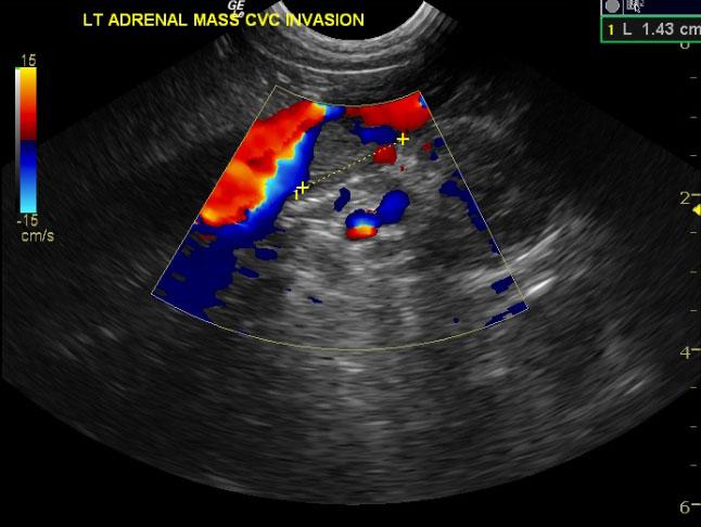
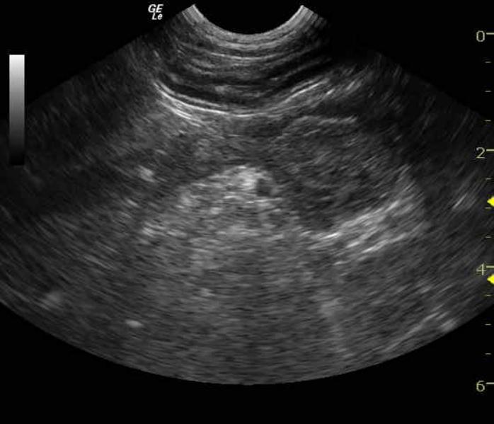
3/2007: The blood chemistry was repeated, and revealed hyperbilirubinemia, hypertriglyceridemia, hyperphosphatemia, and hyperamylasemia. The CBC and TT4 were again within normal limits and the ACTH stimulation again showed elevated cortisol concentrations (pre and post- administration of ACTH.)


DX

Cushing`s disease

Sonographic Differential Diagnosis

Bilateral adrenal enlargement with phrenic thrombus formation. Bilateral adrenal neoplasia is possible but less likely given the long term Cushing`s history of this patient.

Image Interpretation

The adrenal glands presented bilateral uniform hypertrophy for this size patient. Contour was swollen yet uniform. The left adrenal gland was enlarged at 3.2 x 2cm with mottled mixed hypoechoic heterogeneous appearance with echogenic presence of tissue or clot within the phrenic vein and slight impingement into the vena cava. The right adrenal gland was enlarged at 3.5 x 2.5 cm with similar presentation of echogenic tissue which appeared to occupy the phrenic vein and invade approximately 1.3 cm into the caudal vena cava. This is either tumor encroachment or in situ build up of thrombus, which may be due to tissue necrosis from mitotane treatment.

Outcome

The patient was prescribed antithrombotic dose of aspirin due to the suspicion of phrenic thrombosis. The patient remained clinically normal over the next 14 months. No clinical signs of thromboembolic disease were ever noted.

Comments


The adrenal/phrenic vein thrombosis findings were incidental in this patient. When one considers the theoretical model of Virchow’s triad for thrombus formation (hypercoagulable state, endothelial lesion, blood stasis) a Cushingoid dog is a hypercoagulable state. When mitotane necrosis adrenal glands it makes sense that inflammatory events, infarcts, thromboses can occur as the gland is essentially necrosing. Endothelial lesions can naturally occur in an inflammatory state in this case the phrenic veins. This is all theoretical but allowed for reasoning to follow and justify the thrombosis. At the time of this sonogram the plavix was not readily available hence aspirin was administered.


Clinical Differential Diagnosis

Spontaneous hypoadrenocorticism due to adrenal necrosis or idiopathic reaction to mitotane therapy.

Sampling

None taken.

Patient Information

Patient Name : Bailey D
Gender : Female, Spayed
Species : Canine
Type of Imaging : Ultrasound
Status : Complete
Liz Wuz Here : Yes
Code : 07_00028

History

  • Mitotane therapy
  • Pituitary dependent hyperadrenocorticism

Images

LADRbaileydiromaphrenicthrombi2_02062011034417CVCinvasionladrRADRwithphrenicvein

Blood Chemistry

  • Amylase, High
  • Hypertriglyceridemia
  • Phosphorus, High
  • Total Bilirubin, High

Special Testing

  • ACTH Stim Positive for Cushing's
Skip to content