A 3-year-old intact male Jack Russell terrier dog was presented for urinating in the house and not being right. Abnormalities on physical examination were pale mucus membranes and tense abdomen. Anemia and leukocytosis was present on CBC. On survey radiographs a retroperitoneal enlargement causing ventral displacement of the colon was evident.
A 3-year-old intact male Jack Russell terrier dog was presented for urinating in the house and not being right. Abnormalities on physical examination were pale mucus membranes and tense abdomen. Anemia and leukocytosis was present on CBC. On survey radiographs a retroperitoneal enlargement causing ventral displacement of the colon was evident.
Case Study
Hemorrhagic pyelonephritis, causing retroperitoneal bleeding, in a 3 year old intact male Jack Russell
DX
Chronic active, mixed suppurative, hemorrhagic pyelonephritis, associated retroperitoneal hemorrhage
Sonographic Differential Diagnosis
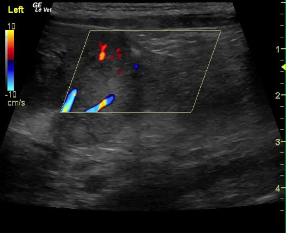
The changes are most consistent with toxic or hemorrhagic nephritis, bilateral renal neoplasia such as lymphoma or other round cell neoplasia, aggressive coagulopathy, or acute, severe pyelonephritis with capsular rupture. The tissue extension deriving from the caudal pole of the left kidney entering into the sublumbar and retroperitoneal space is most consistent with tumor invasion. It appeared to be contiguous with the caudal pole of the left kidney and expanded caudally outside the left kidney itself.
Recommend full coagulation panel in this patient to assess for the coagulopathy that may justify a retroperitoneal hemorrhage. If coagulation times are normal, fine-needle aspirates of the tissue structure in the sublumbar space as well as the cortex in the left kidney would be indicated. Blood pressure measurements, broad spectrum antibiotics, and pain management would all be warranted. Very guarded prognosis.
Image Interpretation
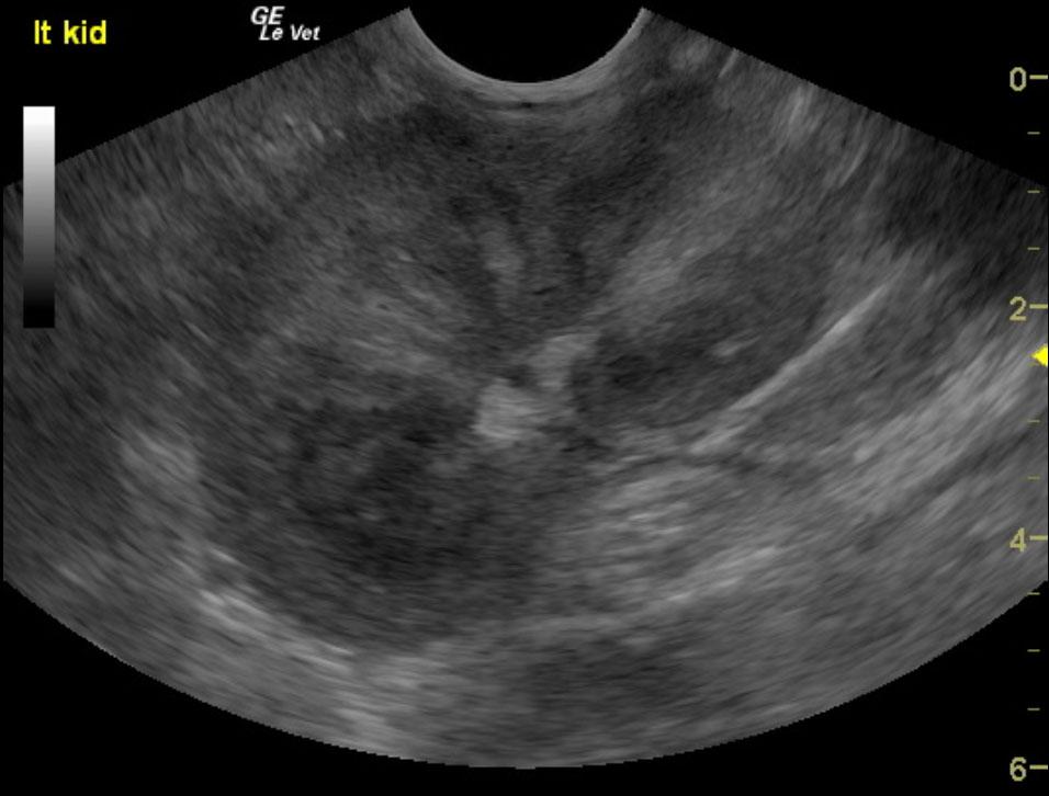
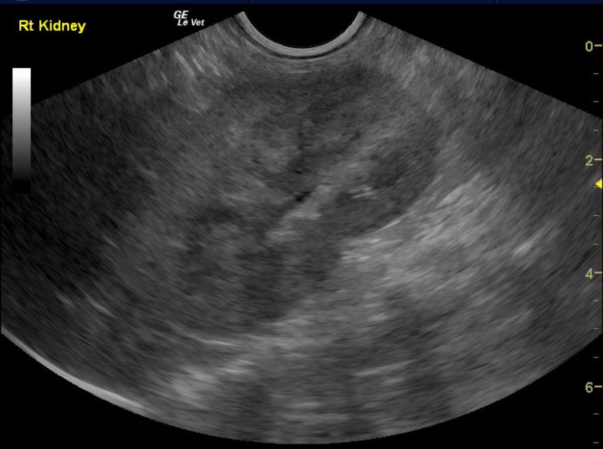
The urinary bladder, trigone and pelvic urethra demonstrated normal wall thicknesses with anechoic urine and normal tone. No uroliths or sediment were visualized. No evidence of inflammatory or neoplastic changes were noted. The ureters were not visible and considered normal. The testicles were uniform without evident pathology. The prostate demonstrated only micronodular changes, and measured 2.77 cm in width. The kidneys in this patient were bilaterally swollen and hypervascular, which is consistent with aggressive, acute disease. The right kidney measured 6.15 x 3.15 cm. There was a complete lack of corticomedullary definition with hypertrophy, micronodular cortical changes and concurrent pyelectasia. Pyelectasia in the right kidney measured 0.25 cm. The patient is on fluid therapy, so the pyelectasia may be caused by fluid and diuresis. Perinephric inflammatory pattern was noted in the surrounding fat. Retroperitoneal space on the left side contained a hypoechoic mass type structure, blood clot or tumor tissue invasion within the sublumbar space that extended from the caudal pole of the left kidney to the level of the urinary bladder. The left kidney measured 5.9 x 3.98 cm with complete loss of the medullary structure, hypervascular cortex and loss of definition at the corticomedullary junction.
Outcome
The patient was euthanized.
Clinical Differential Diagnosis
Renal pathology – trauma, hemorrhage, neoplasia, abscess, acute renal failure, hydronephrosis, ureteral rupture. Abdominal mass effect – neoplasia, abscess, granuloma.
Sampling
After the patient was euthanized, samples from the kidneys were submitted for histopathology. Microscopic findings revealed severe, chronic active, mixed suppurative, hemorrhagic pyelonephritis with associated retroperitoneal hemorrhage, left and right kidneys. These findings are consistent with chronic bacterial infection.
Video
Patient Information
Patient Name :
Rambo T
Gender :
Male, Intact
Species :
Canine
Type of Imaging : Ultrasound
Status :
Complete
Liz Wuz Here :
Yes
Code :
06_00092
Clinical Signs
- "Not Doing Right"
- Inappropriate Urination
Exam Finding
- Pale Mucous Membranes
- Tense Abdomen
CBC
- RBC, Low
- WBC, High
Clinical Signs
- "Not Doing Right"
- Inappropriate Urination


