A 12 year old FS mixed breed dog was presented for chronic intermittent vomiting and decreased appetite (will only eat a liquefied diet.) Additional history was right-sided coxo-femoral luxation. Abnormalities on CBC and serum biochemistry were leukocytosis with a left shift, elevated BUN, elevatedALP, and elevated ALT. On survey radiographs an opacity in the cranial abdomen was evident.
A 12 year old FS mixed breed dog was presented for chronic intermittent vomiting and decreased appetite (will only eat a liquefied diet.) Additional history was right-sided coxo-femoral luxation. Abnormalities on CBC and serum biochemistry were leukocytosis with a left shift, elevated BUN, elevatedALP, and elevated ALT. On survey radiographs an opacity in the cranial abdomen was evident.
Case Study
Duodenal infiltration, suspect neoplasia 12 Year Old FS mixed breed dog
Sonographic Differential Diagnosis
Duodenal infiltrative pattern, suspected duodenal lymphoma or carcinoma with a possibility of aggressive inflammatory disease such as transmural bacterial penetration of bowel infarction secondary post hepatic obstruction with cholecystitis. Recommend fine-needle aspirates of the duodenal wall in this patient avoiding the lumen in order to assess for monocellular versus infiltrate. Underlying lymphoma or carcinoma would be primarily suspected. If mixed inflammation is present then complicated inflammatory bowel disease would be likely. However, fine-needle aspirates would be appropriate of the pancreas. The hope is that adequate medical therapy would allow for clinical improvement if surgery is not a possibility. Surgery would involve resection of the large portion of the upper duodenum. Billroth surgery would likely not be necessary; however, resection up to the pyloric outflow would be necessary. A duodenostomy of the common bile duct would likely also be necessary as well as transposition of the primary pancreatic duct. Guarded to poor prognosis.
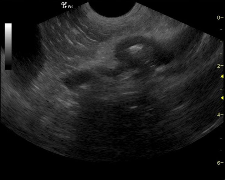
Image Interpretation
The gastrointestinal tract presented pyloric and duodenal wall thickening was surrounded by peri-serosal fat inflammation, which is diffusely hyperechoic. The wall of the duodenum that appeared affected measured 0.72 cm from serosa to mucosa with approximately 1.7 cm in width and 7.0 cm in length. Loss of mural detail was present. The neoplastic criteria was met in this patient. However, underlying complicated inflammatory disease is possible. Fine-needle aspirates of the upper duodenal wall would be recommended to assess any infiltrate that would suggest neoplasia such as lymphoma or carcinoma.
DX
Outcome
The patient was euthanized.
Clinical Differential Diagnosis
Stomach/duodenum – neoplasia, foreign body, IBD, focal perforation, granulomatous gastritis, dietary hypersensitivity Pancreas – pancreatitis, neoplasia Gall bladder – cholecystitis, mucocele, bile duct obstruction
Sampling
None
Video
Patient Information
History
- Decreased appetite
- Vomiting
Images


Blood Chemistry
- Alkaline Phosphatase (SAP), High
- ALT (SGPT), High
- BUN high
CBC
- Left Shift
- WBC, Low