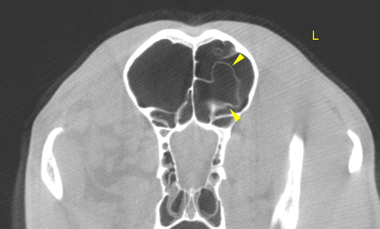
This Labrador mix dog presented with shifting leg lameness of 3 weeks duration,moderately responsive to pain medications. Unilateral nasal bleeding post NSAIDs,(also post trauma) has progressed to constant blood dripping from left nares.
Physical exam: overweight, left nares bleeding, grade 2/4 lameness right front
CBC/Chem: HCT 34%, non-regenerative, PLT 69,000, PT/PTT elevated prior to startng Vit K.