This 11 year old FS Collie dog has a history of megaesophagus and myasthenia gravis
CT of the thorax – There is a soft tissue attenuating cranioventral mass lesion of 4.5 x 3 x 2.5 cm with
well-defined margins. The mass is directly adjacent to the heart but appears to be
separate from the heart. No major vessel or tracheal displacement is noted. The precardial intrathoracic esophagus reveals moderate dilation with gas. There is a hypoattenuating and hypoenhancing nodule of 5 mm diameter within the
right medial liver lobe adjacent to the gallbladder.No dilation
is noted level with the heart and for the caudal thoracic esophagus.
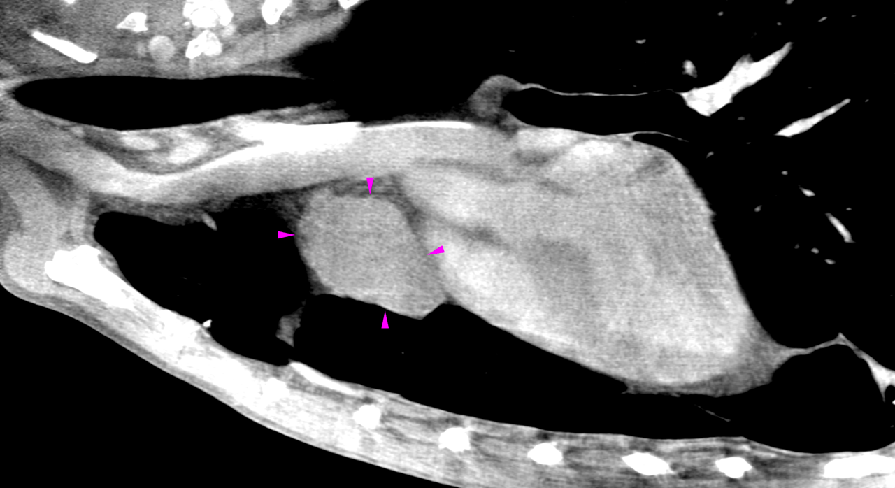
Thymic lymphoma, ectopic thyroid or parathyroid carcinoma and other are all possible,
but by far less likely differential diagnoses. The liver lesion is compatible with a small uncomplicated liver cyst. Benign nodular
hyperplasia or a secondary neoplasia is by far less likely. If further definition is
warranted ultrasound guided sampling would be ideal.