This 10 year old MN Great Pyrenees dog presented with hind end paresis
This 10 year old MN Great Pyrenees dog presented with hind end paresis
This 10 year old MN Great Pyrenees dog presented with hind end paresis
This 10 year old MN Great Pyrenees dog presented with hind end paresis
CT of the lumbar spine, plain series only
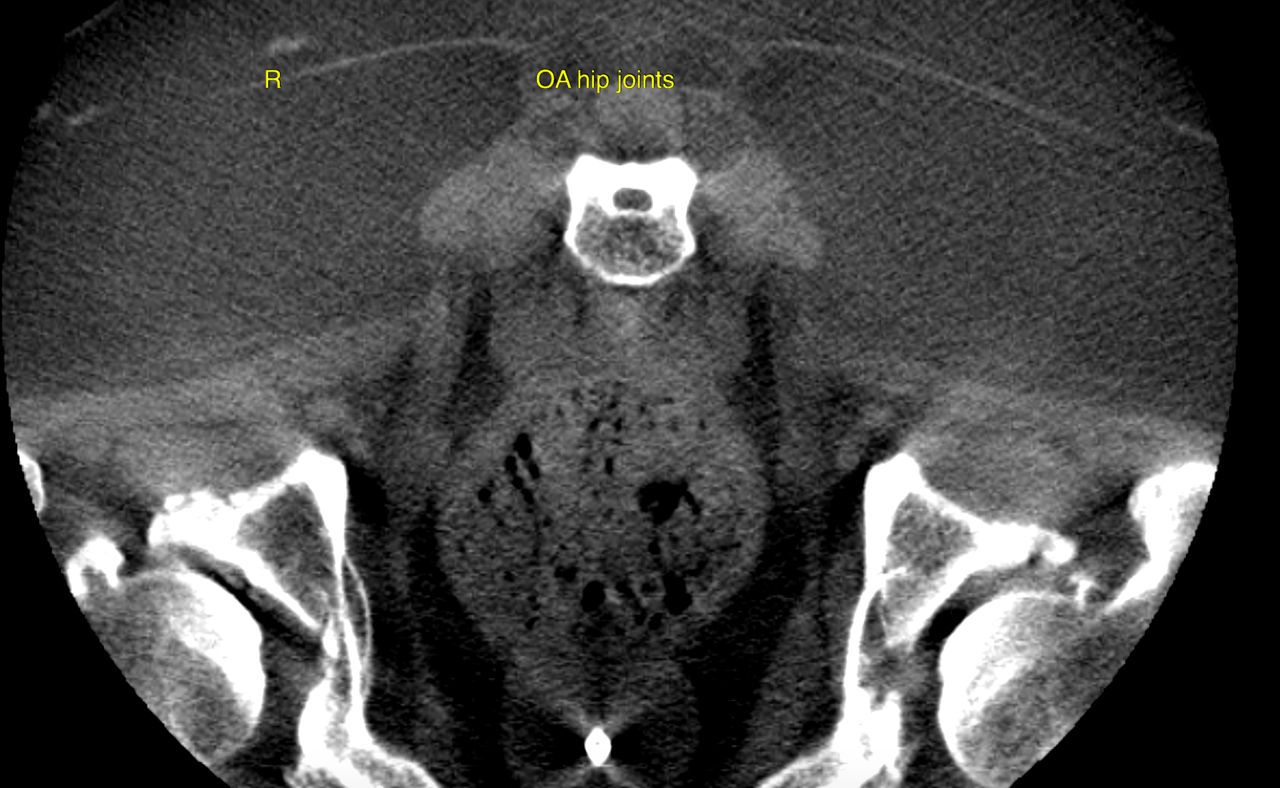
Both hip joints reveal moderate osteoarthritic changes as a function of mild hip
dysplasia.
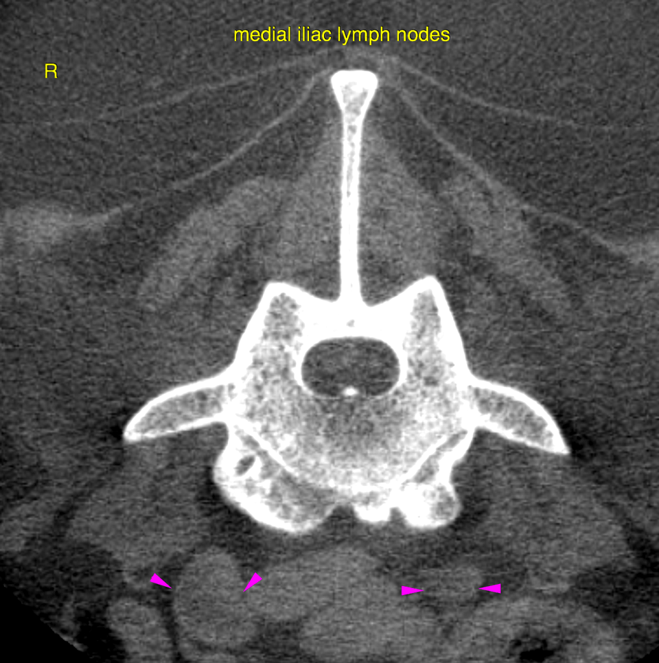
The right medial iliac lymph node presents mild reactive hyperplasia with a benign
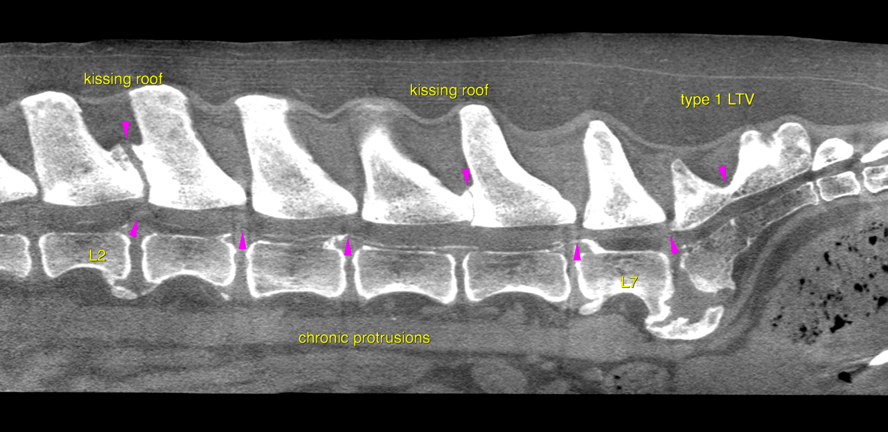
pattern. – A series of chronic disc protrusions of varying degree is seen throughout the lumbar
spine and lumbosacral transition along with overall moderate degenerative changes of
the axial skeleton.
Two locations stand out regarding the severity of changes:
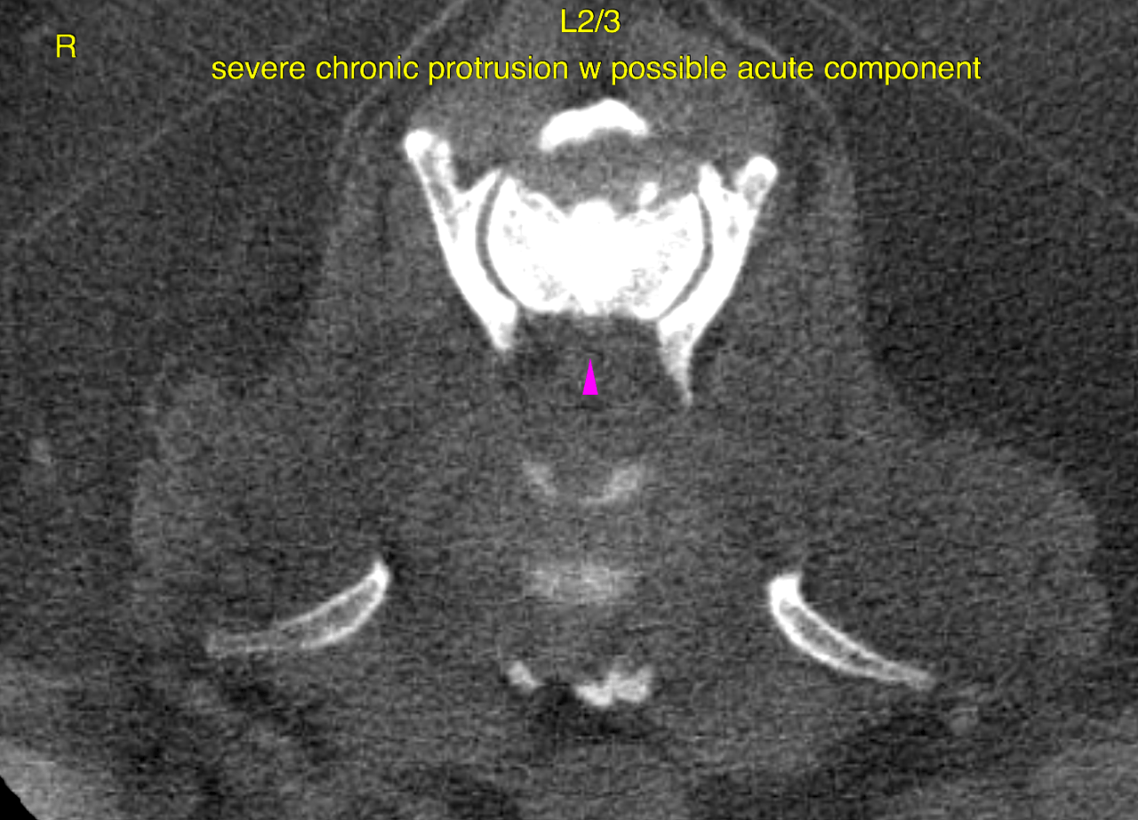
The disc L2/3 reveals a severe protrusion with marked dorsal displacement and
compression of the spinal cord. Moreover soft tissue attenuating compressive material
extends caudally within the ventral epidural space up to the mid of the L3 vertebral
body.
The lumbosacral disc reveals a severe protrusion with complete occupation of the
vertebral canal diameter as well as marked bilateral neuroforaminal narrowing. The
nerve fibres of the cauda equina cannot be delineated against the disc material. There is
a marked spondylosis, spondylarthrosis, demineralization of the sacrum as well as a
long cranial and ventral extension of the sacral roof and a lumbosacral step formation.
The first spinous process has remained unfused (Type 1 lumbosacral transitional
vertebra).
Mild to moderate chronic disc protrusions are seen at L3/4, L4/5, L6/7. The degree of
compressive myelopathy is mild to moderate here respectively. Extensive facet arthropathy and „kissing“ vertebral arches are seen at L2/3 and L5/6.
• Moderate osteoarthritis of both hip joints due to dysplasia
• Unilateral mild medial iliac lymph node hyperplasia of unknown originClinical correlation and neuroanatomic localization to the upper or lower motor neuron
is crucial here to determine the relevant site of compression.