CT of the head and thorax –
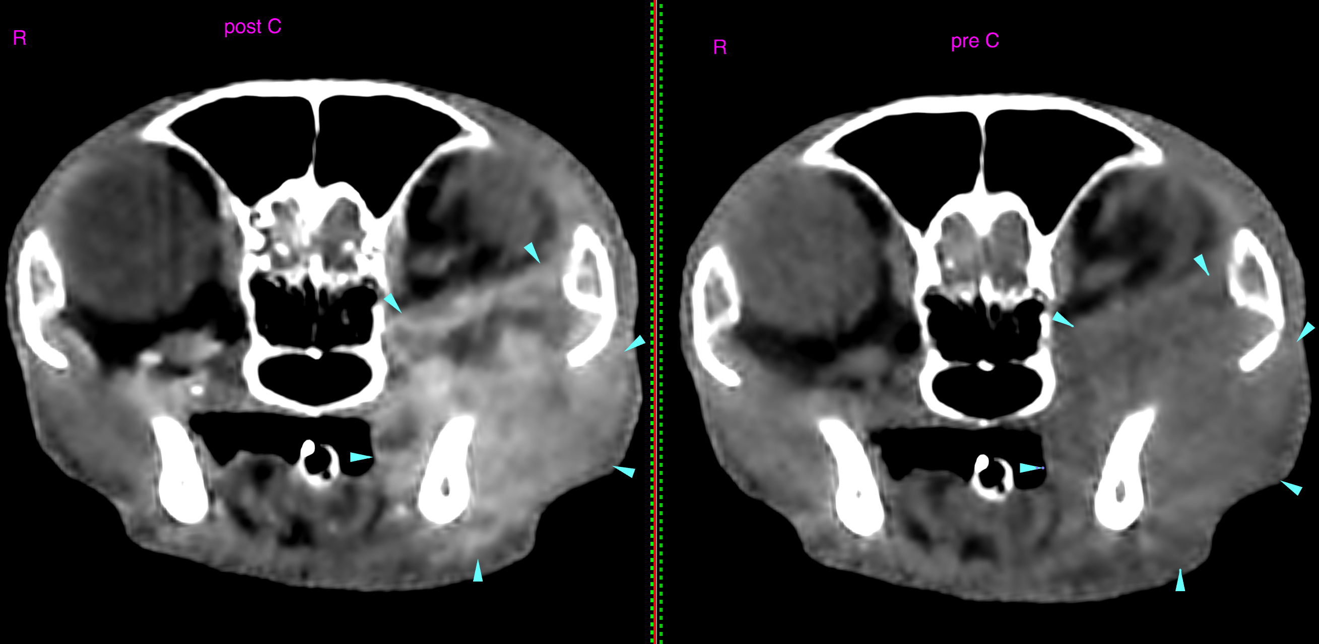
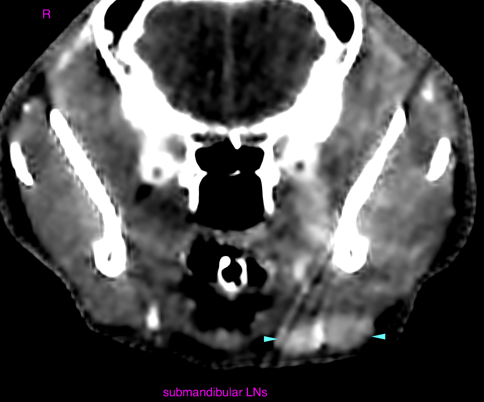
An ill-defined mass effect is noted within the left retrobulbar & retromolar space and masseter region displacing the globe temporodorsally. The lesion shows moderate, non-uniform contrast enhancement with a predominating ring-like pattern. The lesion has a significant mass effect onto the optic nerve, extraocular musculature and oropharynx. The globe is macroscopically not infiltrated. No aggressive osteolysis is seen. The left medial retropharyngeal lymph node and the left submandibular lymph nodes are mildly enlarged with preserved short to long axis ratio and enhancement pattern. Except for generalized periodontal disease with jaw bone atrophy and FOR lesions the dentition does not present abnormalities. An association with the retrobulbar process is not evident