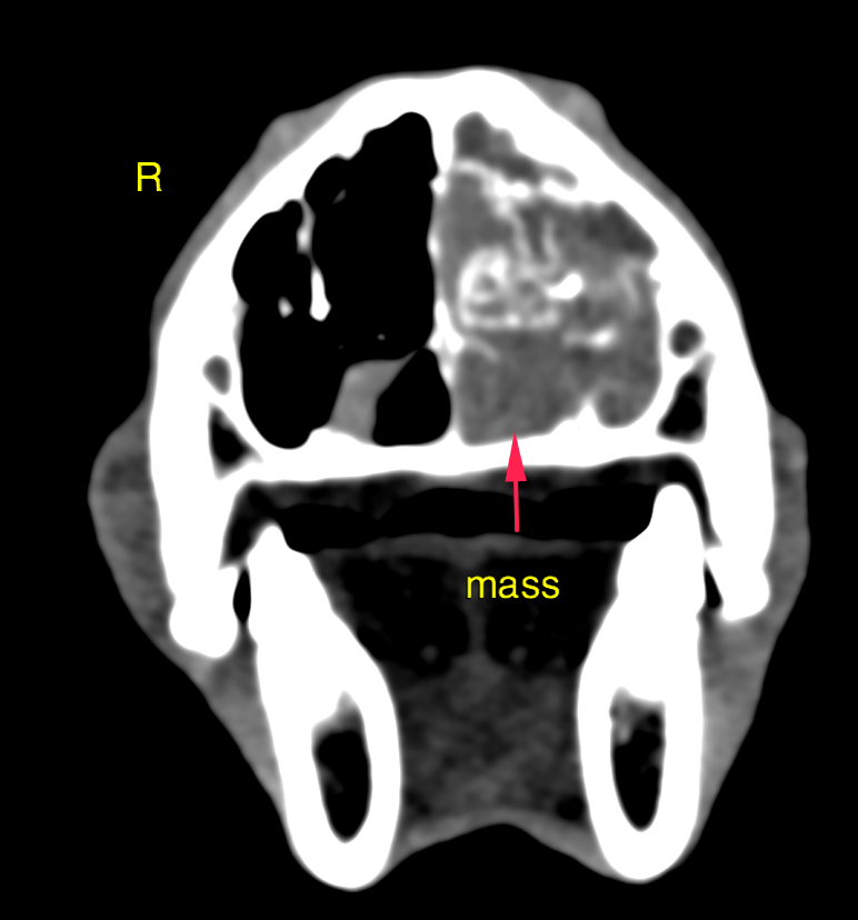
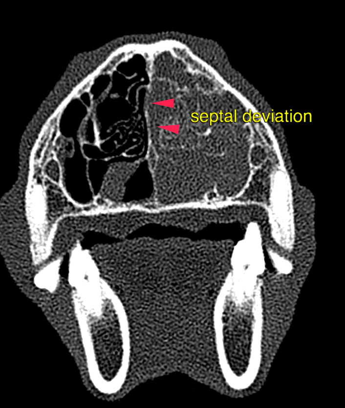
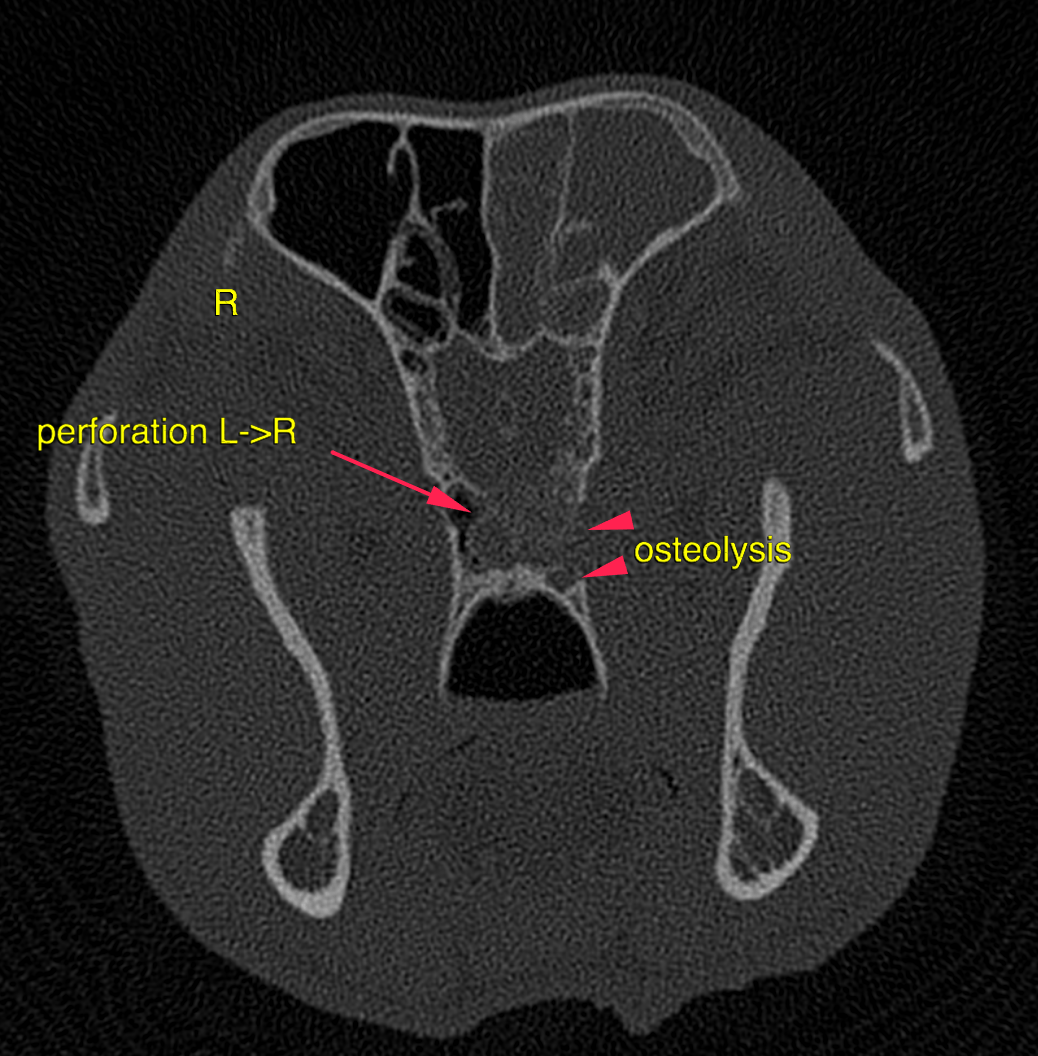
This 12 year old M Husky has a history of chronic intermittent sneezing, reverse sneezing and epistaxis, all of which has been increasing in frequency and severity. Mucoid nasal discharge
This 12 year old M Husky has a history of chronic intermittent sneezing, reverse sneezing and epistaxis, all of which has been increasing in frequency and severity. Mucoid nasal discharge