CT – Portosystemic shunt, micro hepatica and ureteral obstruction in a 4 year old MN Shih Tzu

Case Study

CT – Portosystemic shunt, micro hepatica and ureteral obstruction in a 4 year old MN Shih Tzu

This 4 year old MN Shih Tzu has a history of chronic liver disease and elevated bile acids. Liver biopsy dx vascular anomaly. Intermittent abnormal behavior and hematuria. slighty overweight. Bile acids 164/123

This 4 year old MN Shih Tzu has a history of chronic liver disease and elevated bile acids. Liver biopsy dx vascular anomaly. Intermittent abnormal behavior and hematuria. slighty overweight. Bile acids 164/123

DX

Portosystemic shunt and ureteral obstruction

Image Interpretation

CT of the abdomen – 

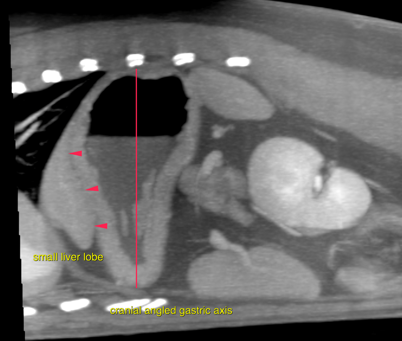
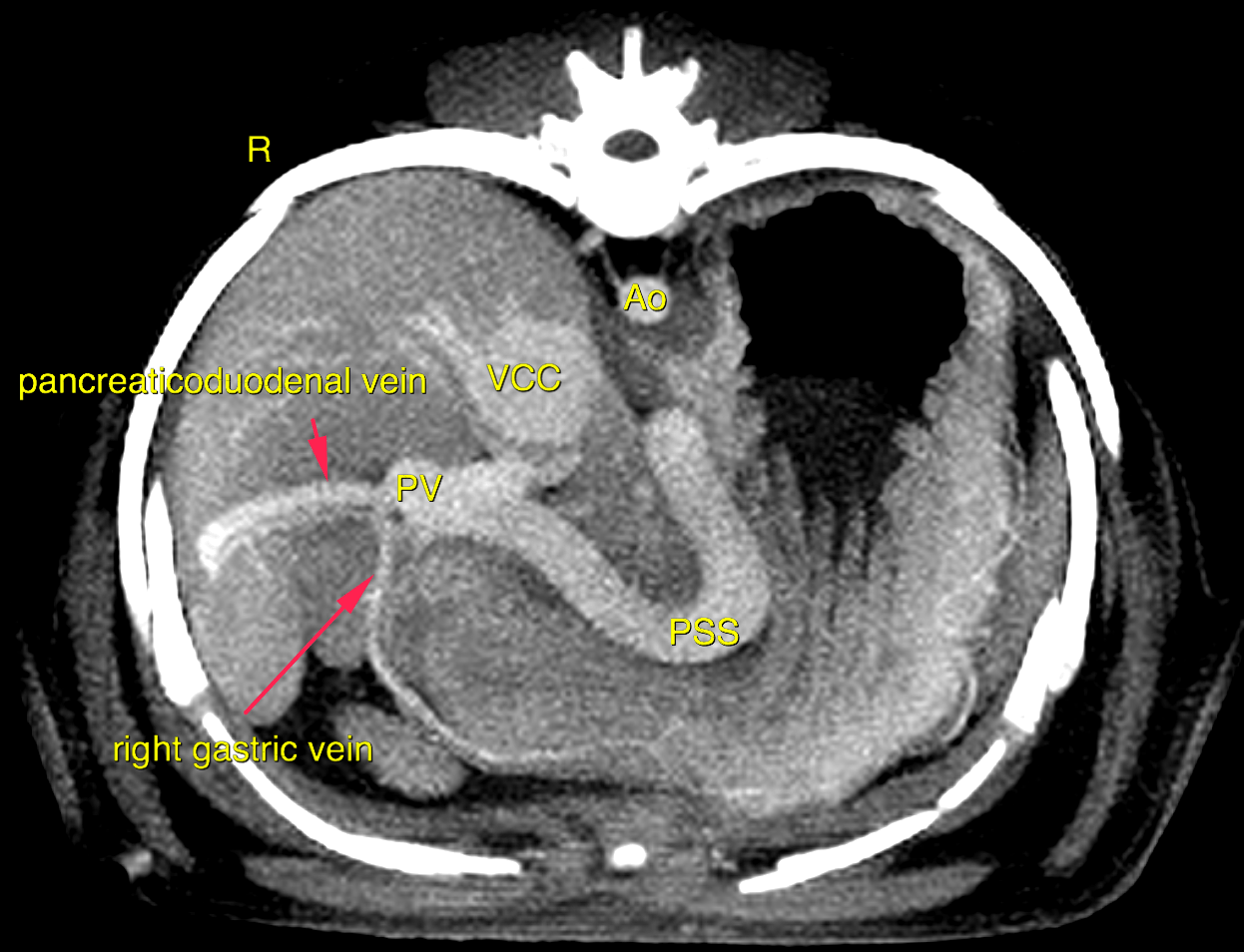
Cranial to the junction of the gastroduodenal vein with the portal vein an aberrant vessel is noted emerging from the left aspect of the portal vein. The aberrant vessel loops along the minor curvature of the stomach and then courses dorsally and caudally to merge with the caudal vena cava from the left side at the level of T11/T12. A small isthmus of the dilated shunt vessel is seen at the junction with the caudal vena cava. Cranial to the aberrant vessel only a rudimentary portal vein is seen. Especially the central and left portal vein branches are miniscule. The liver presents a markedly reduced volume and portal vein branching emphasizing the central and left liver lobes. 

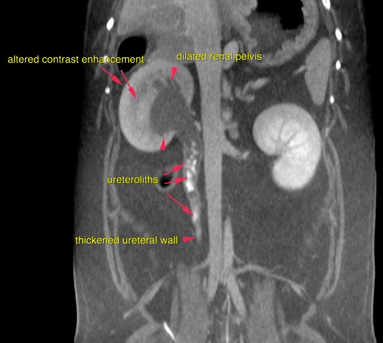
Nonobstructive renal calculi precipitate within the left renal pelvis.  The right kidney presents an altered nephrogram with homogeneous, but reduced contrast enhancement compared to the normal left kidney.  The right renal pelvis is dilated at 2 cm in diameter. The proximal ureter is dilated at 1 cm. At the level L2 to cranial endplate of L4 multiple mineral attenuating objects are noted within the lumen of the right ureter. The distal right ureteral wall is moderately thickened.

Outcome

Single congenital extrahepatic porto-systemic shunt (porto-caval-shunt)
• Microhepatica
• Non-obstructing left-sided renal calculi
• Right sided ureterolithiasis with ureteral obstruction, hydroureter, ureteritis,
pyelectasia and obstructive nephropathy
• Cystolithiasis
The findings confirm the suspected porto-systemic bypass by a single porto-caval
shunt vessel.
Surgical intervention by a slow progressive closure technique (ameroid constrictor,
cellophane banding) is the therapy of choice. Empirical treatment until surgery along
with feeding of a hepatic diet is recommended.
The ureteroliths and cystoliths are most likely related to the porto-systemic metabolic
syndrome that facilitates formation of urate crystals. Interventional (SUB =
subcutaneous ureteral bypass) or surgical (kidney saving ureterotomy – which
unfortunately carries a high risk of postprocedural stricture – versus nephrectomy)
options should be discussed to correct the right ureteral obstruction.

Patient Information

Patient Name : Romeo Gonzalez
Gender : Male, Neutered
Species : Canine
Type of Imaging : Ultrasound
Status : Complete
Liz Wuz Here : Yes
Code : 16_00164

Clinical Signs

  • "Not Doing Right"

History

  • Elevated Bile Acids

Images

bildschirmfoto_2016-09-13_um_10bildschirmfoto_2016-09-13_um_10bildschirmfoto_2016-09-13_um_10

Clinical Signs

  • "Not Doing Right"

Urinalysi

  • Blood Present
Skip to content