This 2 year old German Shorthair Pointer mix has a history of urinary incontinence and hooded vulva. R/O ectopic ureter
CT of the abdomen –
Both kidneys present within normal limits for size, shape and architecture. The nephroand pyelogram phase is bilaterally symmetric and considered normal. The ureters are not dilated and show a physiological course. Both ureters terminate approximately 1.5 and 1 cm cranial to the bladder neck within the urinary bladder. The right vesicourethral junction (VUJ) is slightly cranial to the left which is considered within normal limits. The position of the urinary bladder and urinary bladder neck is relatively far caudal within the pelvis canal. No contrast medium is seen within the urethra in the early and late post contrast phases.
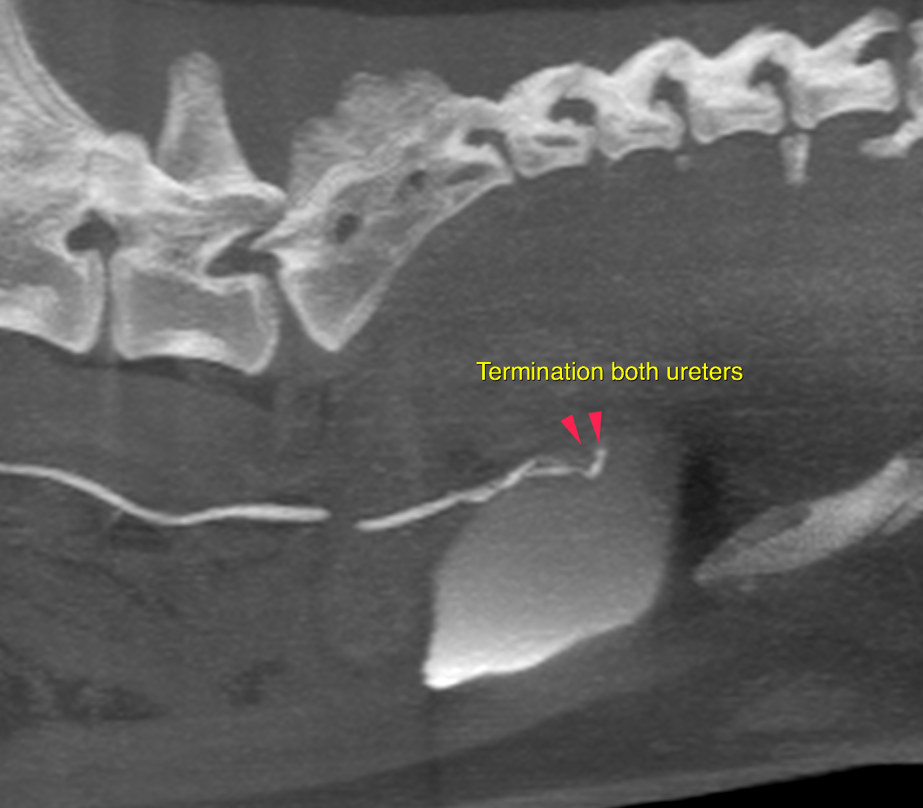
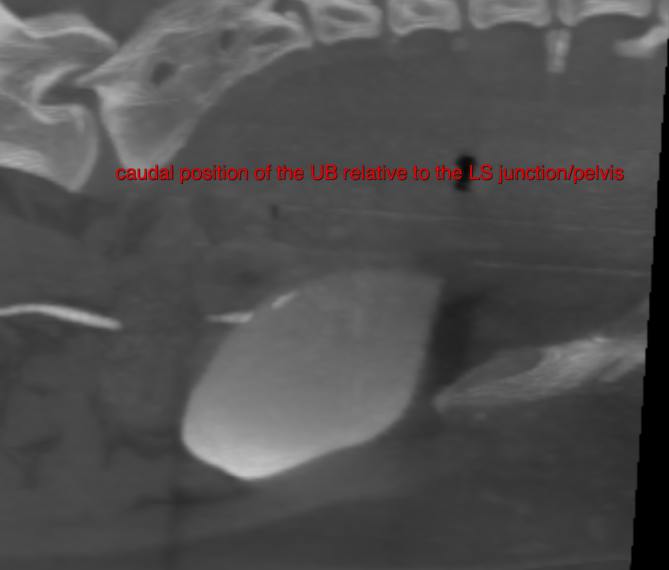
Pelvic bladder with relative caudal position
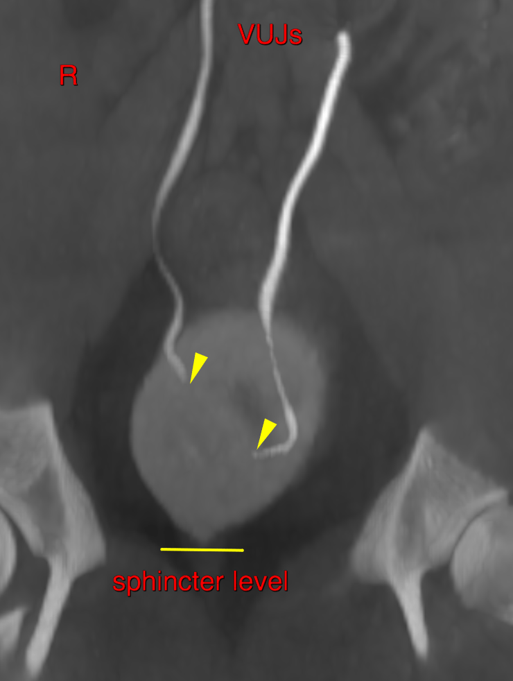
• No evidence of ureteral ectopic
Theoretically an intramural ectopic ureter cannot be ruled out entirely. In case of strong clinical suspicion further workup by means of cystoscopy may be considered. However, this is unlikely.
The significance of the pelvic bladder remains undetermined. Many dogs with the
same relative position of the urinary bladder do not present urinary incontinence. Other
underlying causes – such as urethrocele, urinary bladder/lower sphincter dyssynergia,
behavioral problems, juvenile incontinence, PU/PD – need to be considered as well.
Also note that dogs that are spayed early at an age of < 3 months are known to have an
incidence of incontinence of 12 % and dogs spayed at an age > 3 months of 5%*.
Consider a therapy with ephedrine as first choice medication.