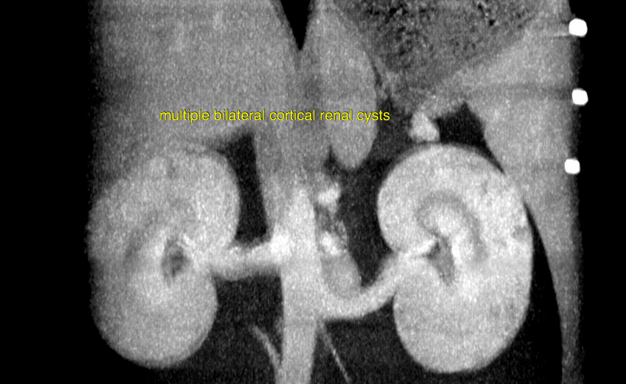
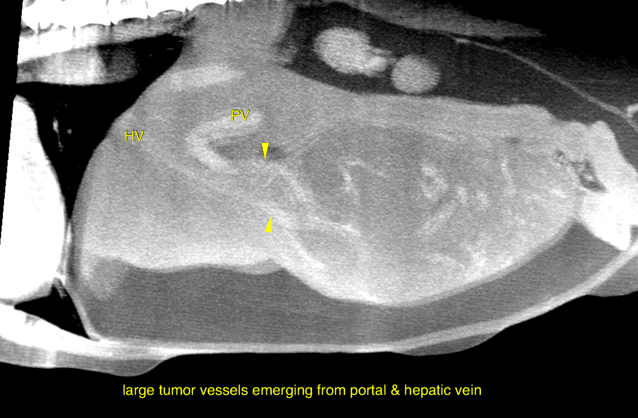
This 14 year old FS Rat Terrier dog presented for a palpable liver mass, confirmed with ultrasound. CT to determine if resectable.
This 14 year old FS Rat Terrier dog presented for a palpable liver mass, confirmed with ultrasound. CT to determine if resectable.