CT of the head and cranial thorax –
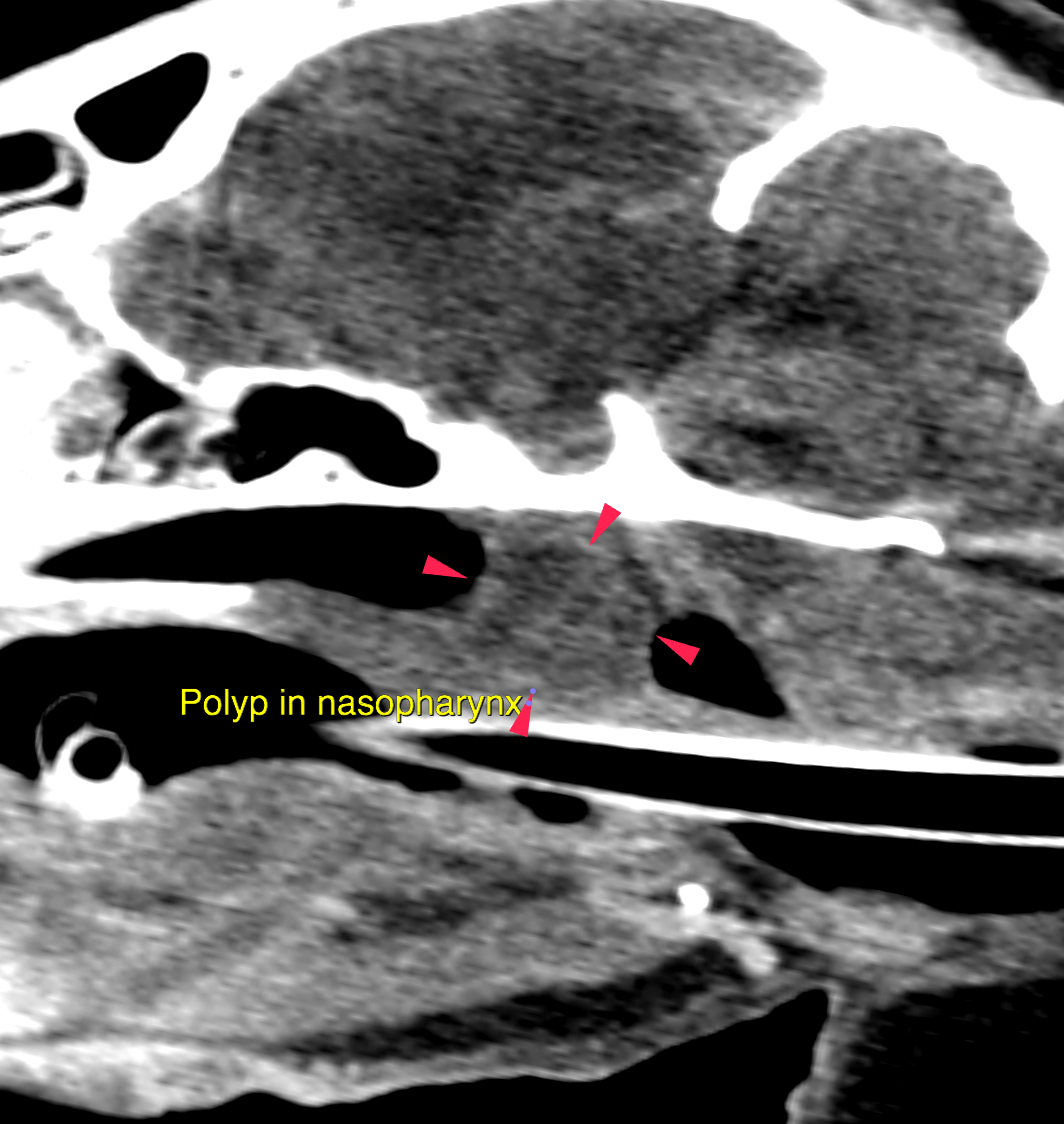
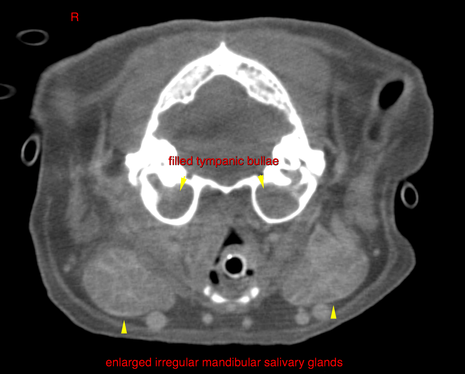
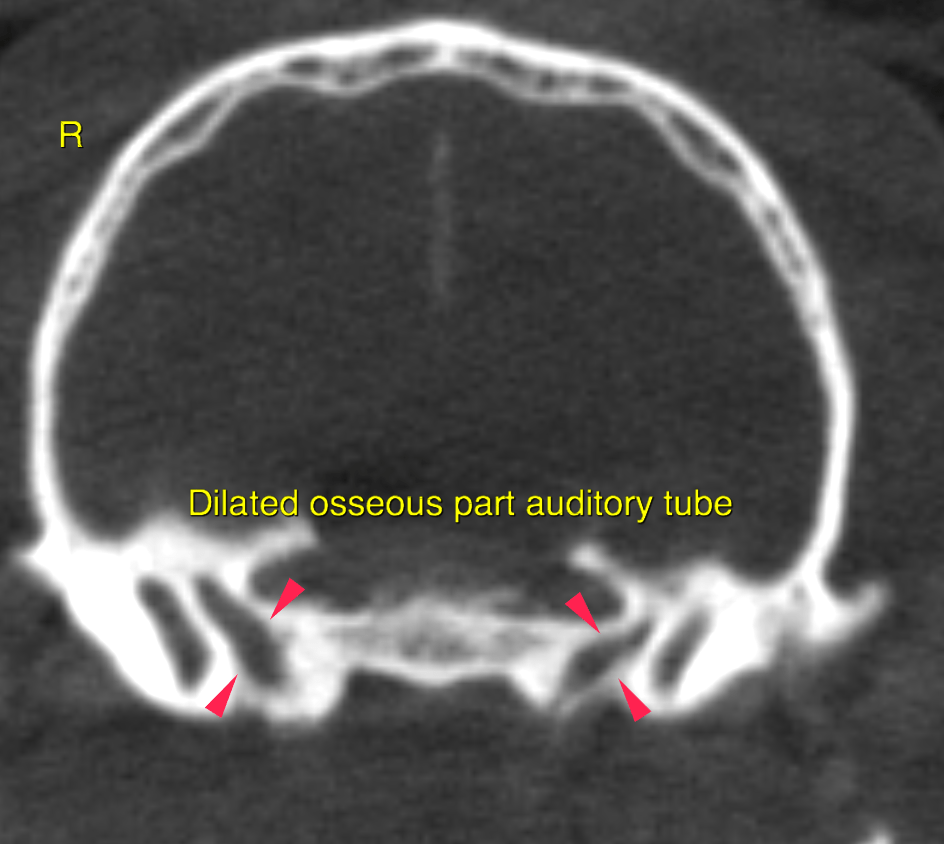
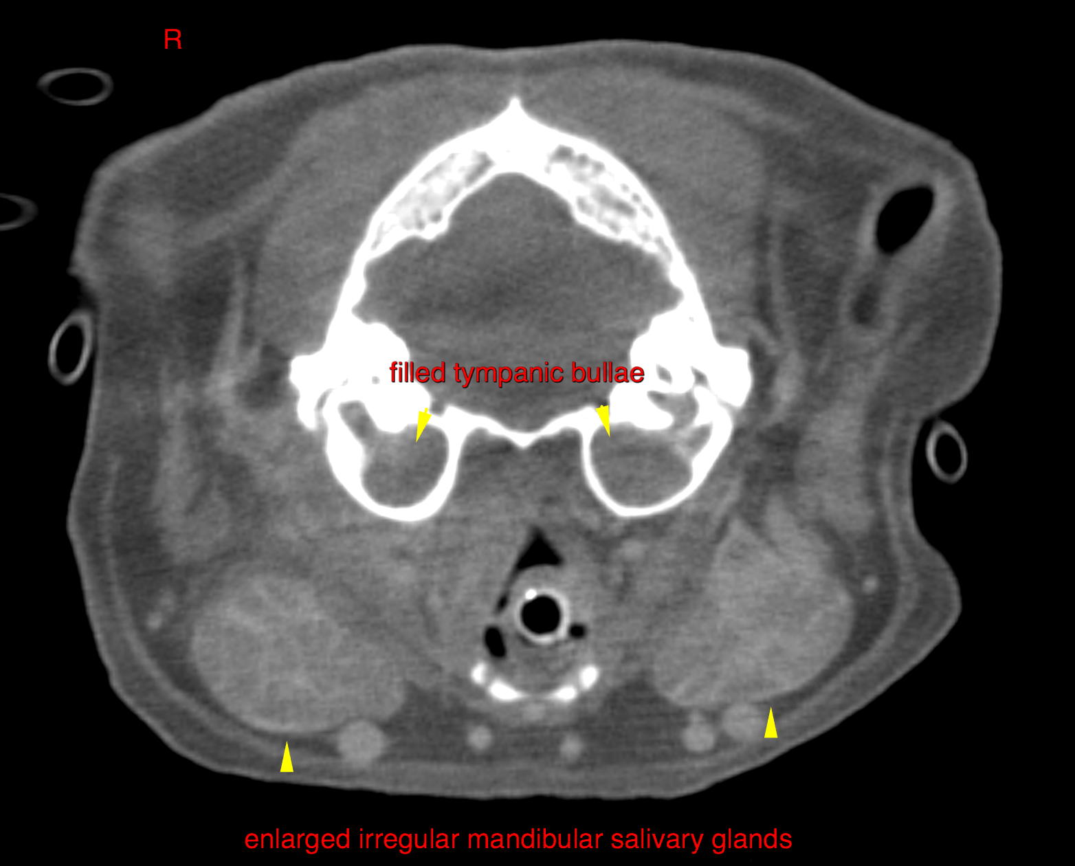
Both tympanic bullae are completely filled with homogeneous soft tissue attenuating material, which can be traced through the auditory tubes to the nasopharynx. The nasopharynx is completely obliterated by a 1 x 1.2 x 1.5 cm ovoid soft tissue mass with faint peripheral contrast enhancement and a mass effect onto the soft palate. Moderate symmetric enlargement of both mandibular salivary glands. An intracranial extraaxial, uniformly enhancing mass lesion of 8 mm height with broad attachment to the bone surface is seen in the midline within the sella region. The mass causes centrifugal displacement of the surrounding brain parenchyma. The caudal compartment of the left cranial lung lobe presents completely soft tissue attenuating and the volume is markedly reduced with an indistinct air bronchogram.


