CT – Nasal soft tissue neoplasia with osteolysis, rhinitis and sinusitis in a 10 year old FS Boxer mix

Case Study

CT – Nasal soft tissue neoplasia with osteolysis, rhinitis and sinusitis in a 10 year old FS Boxer mix

This 10 year old FS Boxer mix dog presented with a bloody nasal discharge

This 10 year old FS Boxer mix dog presented with a bloody nasal discharge

Image Interpretation

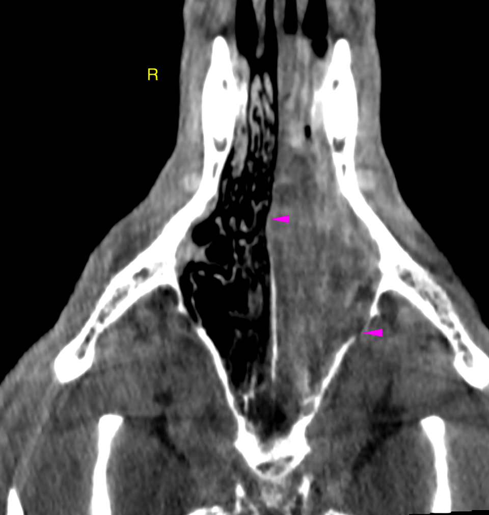
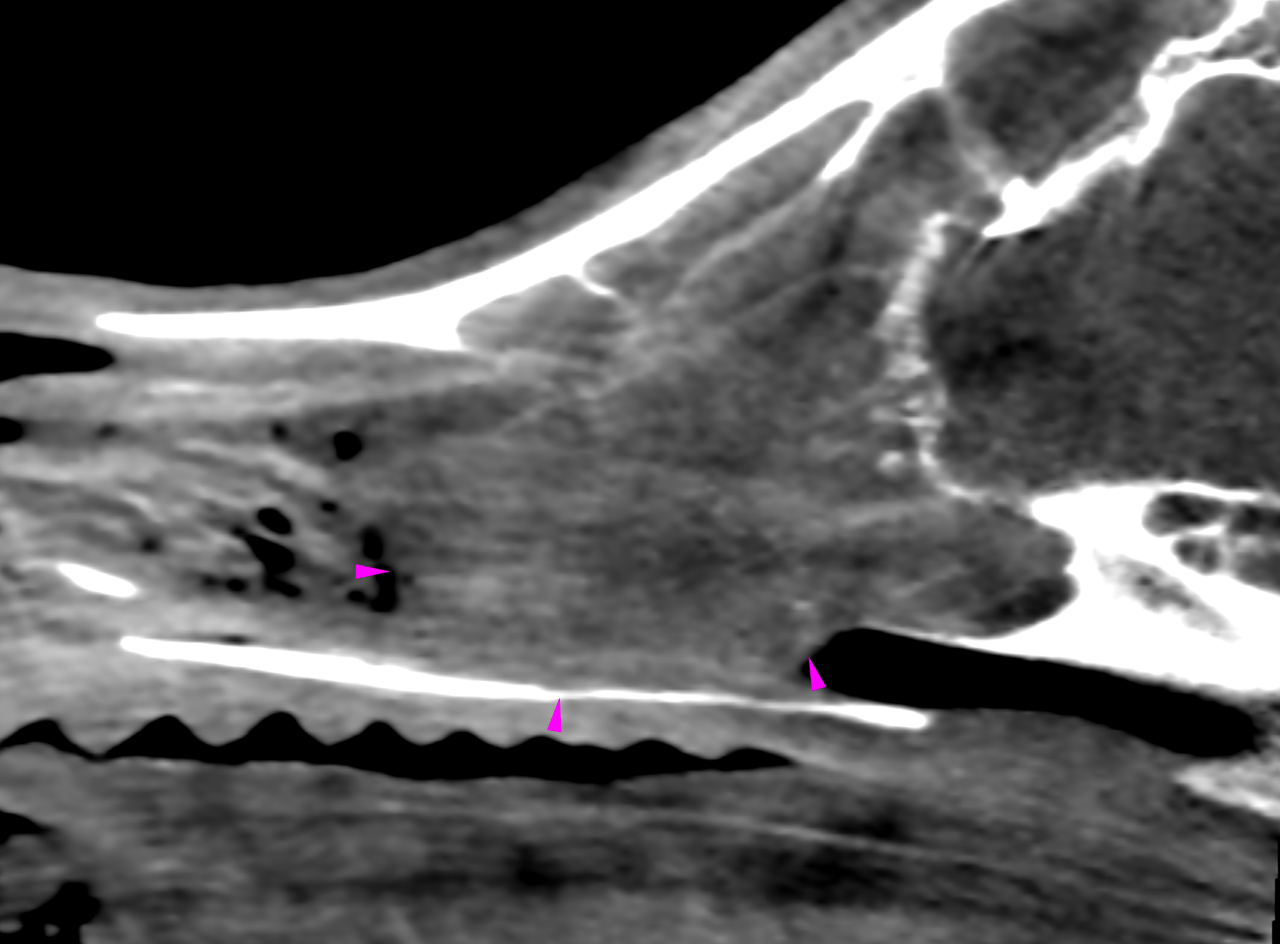
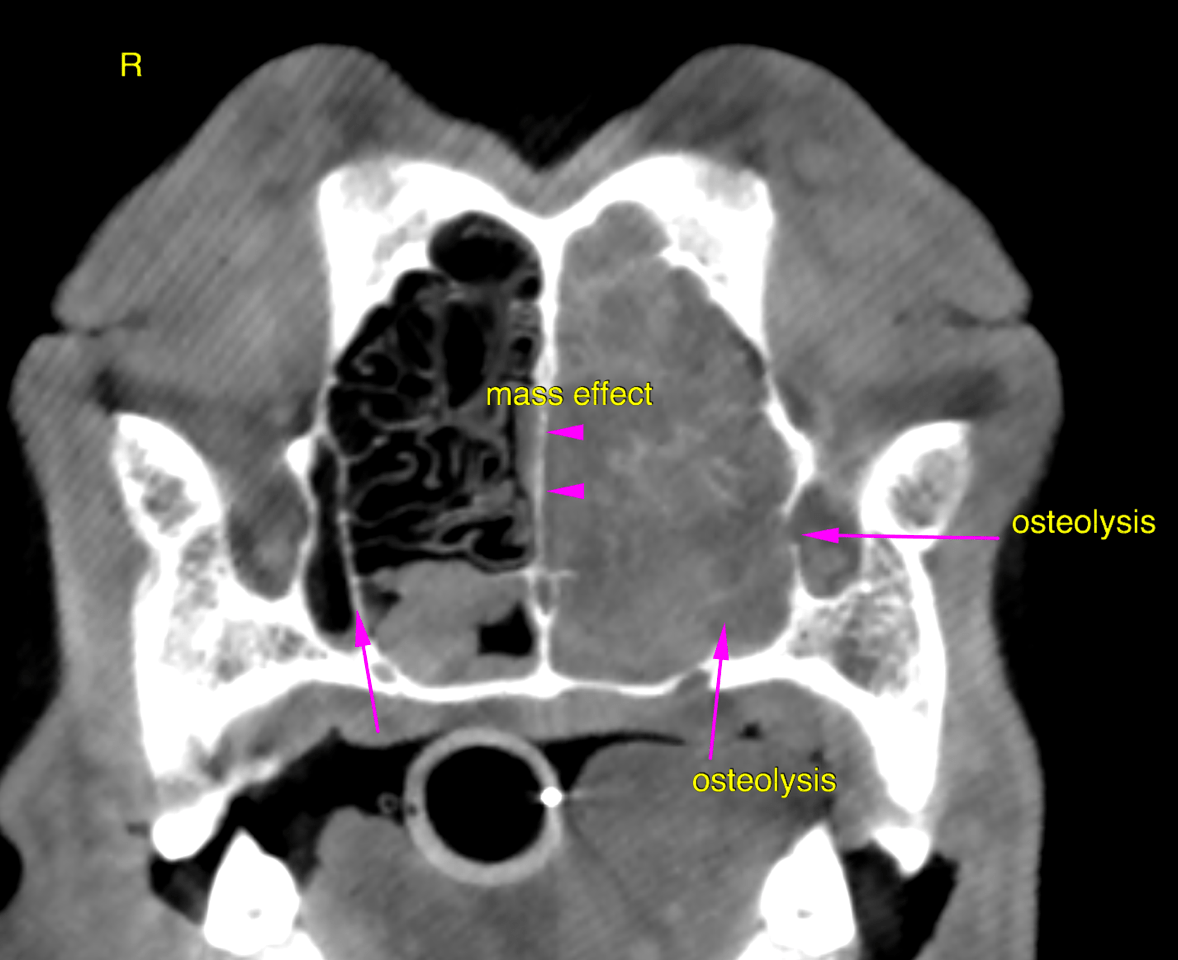
CT of the head – The computed tomography of the head reveals a soft tissue attenuating and nonuniformly
enhancing expansile mass lesion of 4 x 3.5 x 2.5 cm within the mid and
caudal thirds of the left nasal cavity. The mass presents infiltrative growth with
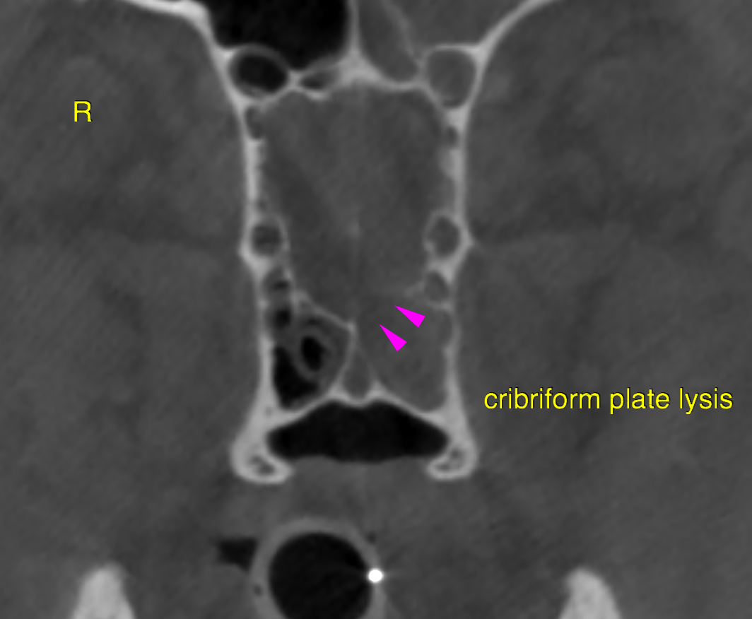
extensive nasal turbinate destruction as well as multifocal osteolysis of the nasal,
maxillary, palatinal, vomer bone, bony orbita and cribriform plate. There is no
intracranial extension.
The left frontal sinus contains fluid attenuating secretions. The left nasal passage is
completely obstructed.
DX

The computed tomographic findings are compatible with a malignant left-sided expansile nasal soft tissue neoplasia with secondary aggressive osteolysis and secretory rhinitis and sinusitis

Outcome

Possible differentials include lymphosarcoma, adenocarcinoma, squamous cell
carcinoma, transitional cell carcinoma, fibrosarcoma and other.
Rhinoscopy and biopsies of the mass should be obtained for further definition. Complete surgical resection is not an option owing to the extent of the lesion. Consider
full tumor staging and radiation therapy and/or chemotherapy with or without
cytoreductive surgery depending on the histopathological results as treatment options

Patient Information

Patient Name : Coco Chanel Hourigan-Thomas/Bluegrass
Gender : Female, Spayed
Species : Canine
Type of Imaging : Ultrasound
Status : Complete
Liz Wuz Here : Yes
Code : 16_00021

Clinical Signs

  • Nasal discharge bloody

Images

bildschirmfoto_2016-05-24_um_08bildschirmfoto_2016-05-24_um_08bildschirmfoto_2016-05-24_um_09bildschirmfoto_2016-05-24_um_09

Clinical Signs

  • Nasal discharge bloody