CT – Multifocal neoplastic mass lesions of the lung with obstruction of the main stem bronchus in a 9 year old MN Welsh Corgi

Case Study

CT – Multifocal neoplastic mass lesions of the lung with obstruction of the main stem bronchus in a 9 year old MN Welsh Corgi

This 9 year old MN Welsh Corgi presented with an extraluminal mass obstructing the left mainstem bronchus.

This 9 year old MN Welsh Corgi presented with an extraluminal mass obstructing the left mainstem bronchus.

DX

• Multifocal bronchocentric and peribronchial mass lesions of the left lung with obstruction of the left main stem bronchus • Severe interstitial and bronchial infiltrate of the left lung • Resorption atelectasis left lung with compensatory hyperinflation of the right lung • Mediastinal lymphadenopathy involving the cranial and tracheobronchial lymph nodes

Image Interpretation

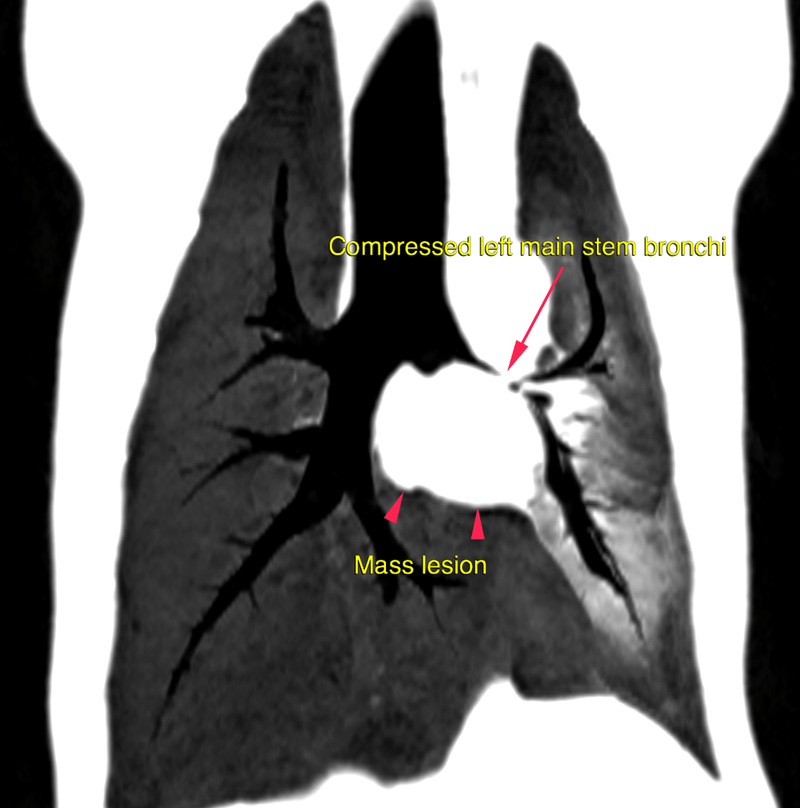
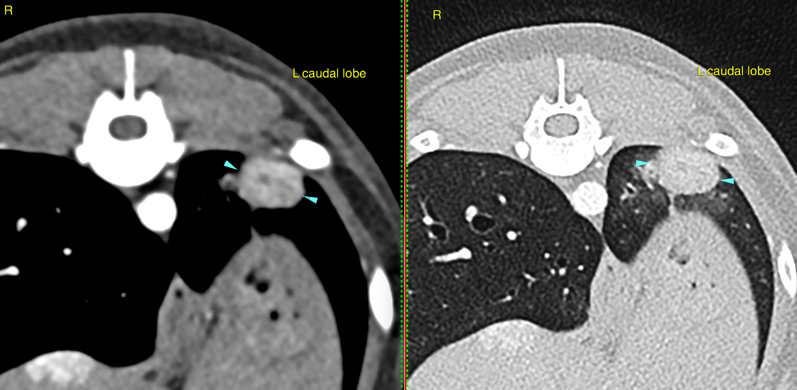
CT of the thorax – A bronchocentric, ill-defined and non-uniformly enhancing soft tissue mass lesion of 2.6 x 3 x 3.6 cm is seen circumferential to the left main stem bronchus. A severe mass effect on the left main stem bronchus is noted and causes focal obliteration of the bronchial lumen. The tracheal wall is moderately thickened at the level of the carina and cannot be delineated from the mass lesion. A second ovoid well-delineated soft tissue mass lesion is visible within the most caudodorsal aspect of the left caudal lung lobe and measures 1.1 x 1.5 x 1.1 cm.  A third small & nodular soft tissue attenuating lesion of 0.2 x 0.4 x 0.3 cm is seen within the lateral subpleural aspect of the caudal compartment of the left cranial lung lobe. 

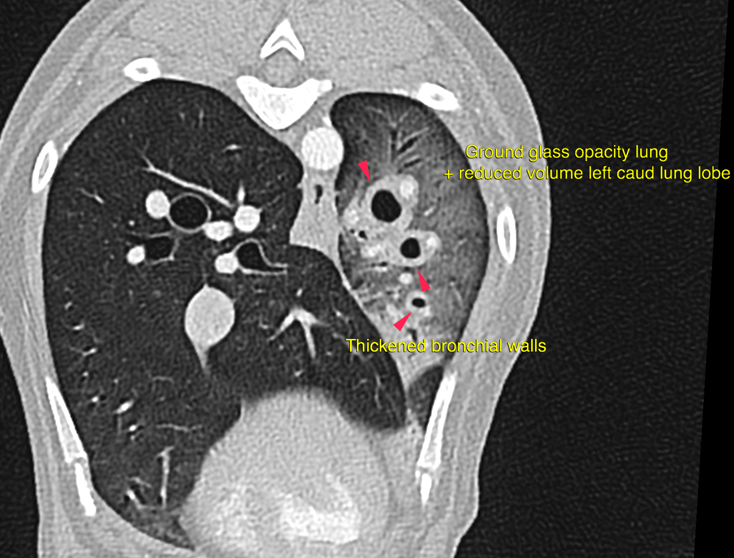
The remainder of the left lung presents an overall decrease in with ground glass opacity. The ventral aspect of the caudal compartment of the left cranial lung lobe reveals a focal alveolar infiltrate.

Outcome

The findings are compatible with a bronchial cell carcinoma with complete infiltration of all bronchial walls of the left lung lobes and metastatic spread to the lung parenchyma and the tributary lymph nodes. The thickening of the tracheal wall is highly suspicious for local invasion of the tracheal wall.
Due to the severe mass effect of the circumferential mass lesion at the left main stem bronchus there is complete bronchial obliteration with secondary resorption atelectasis.

Patient Information

Patient Name : Corky/VetMed Consultants
Gender : Male, Neutered
Species : Canine
Type of Imaging : Ultrasound
Status : Complete
Liz Wuz Here : Yes
Code : 16_00106

Exam Finding

  • Masses

Images

bildschirmfoto_2016-08-19_um_09bildschirmfoto_2016-08-19_um_09bildschirmfoto_2016-08-19_um_09bildschirmfoto_2016-08-19_um_10
Skip to content