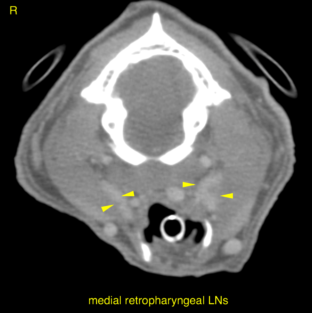
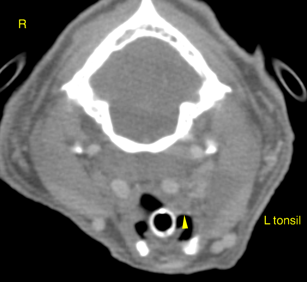
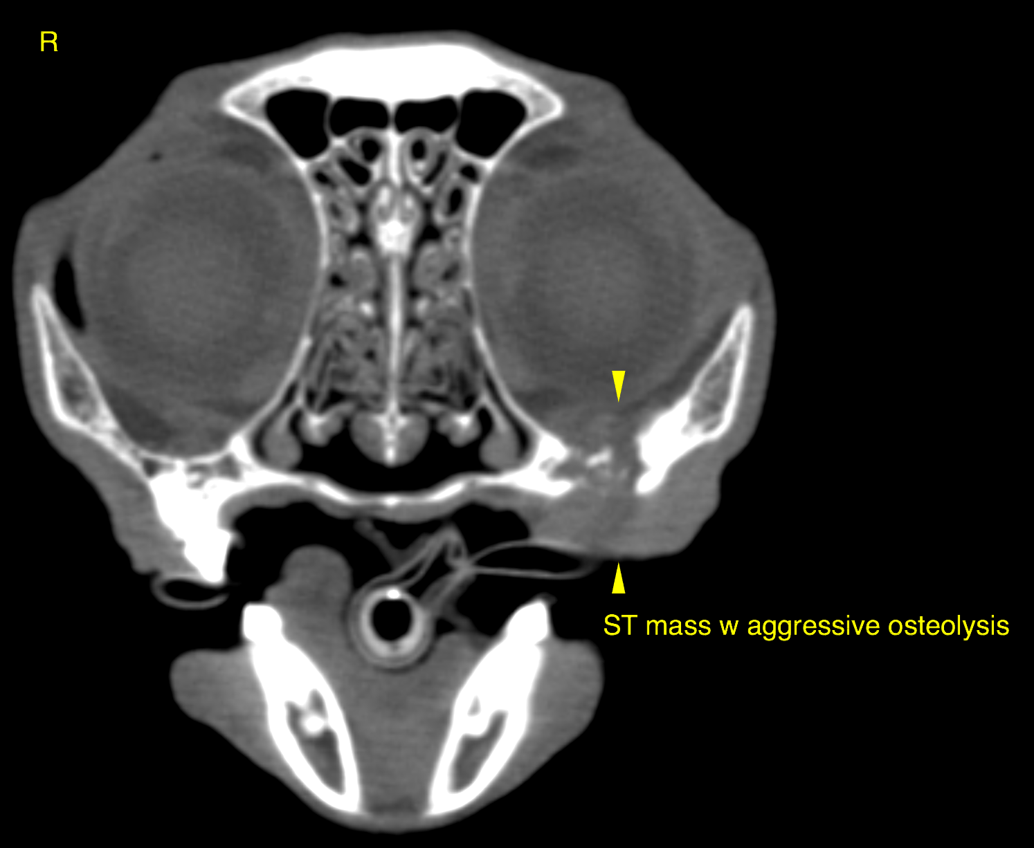
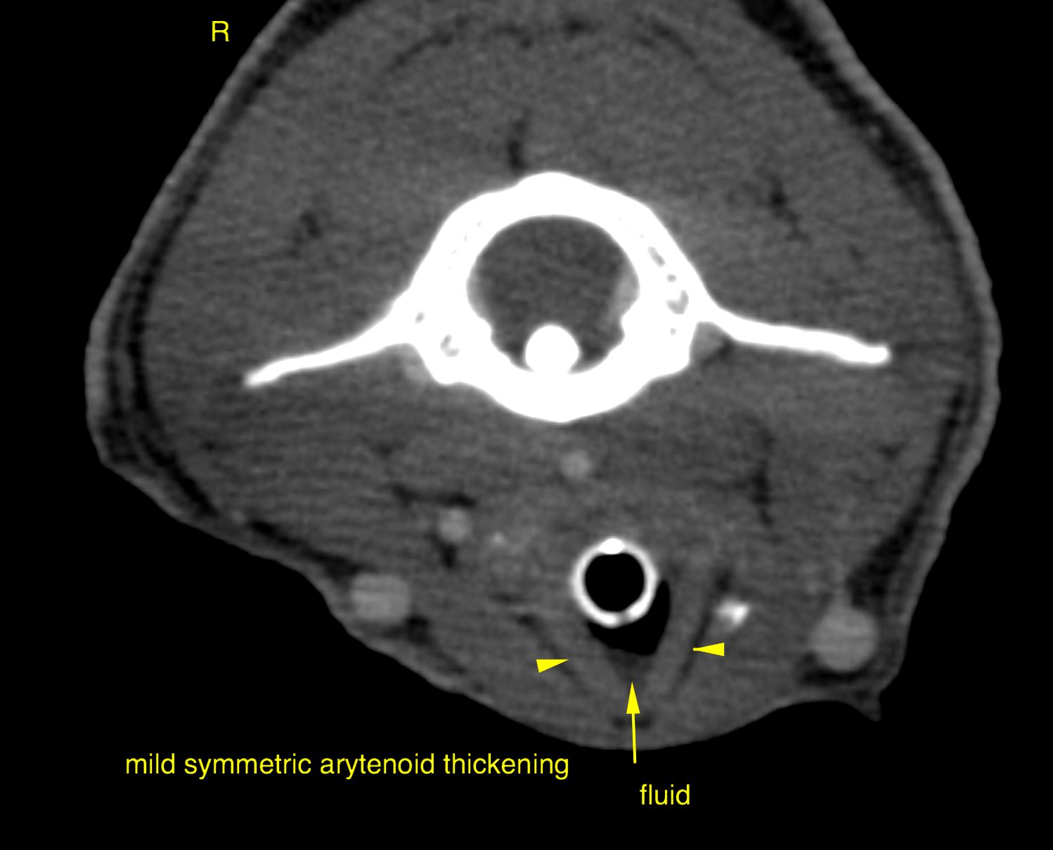
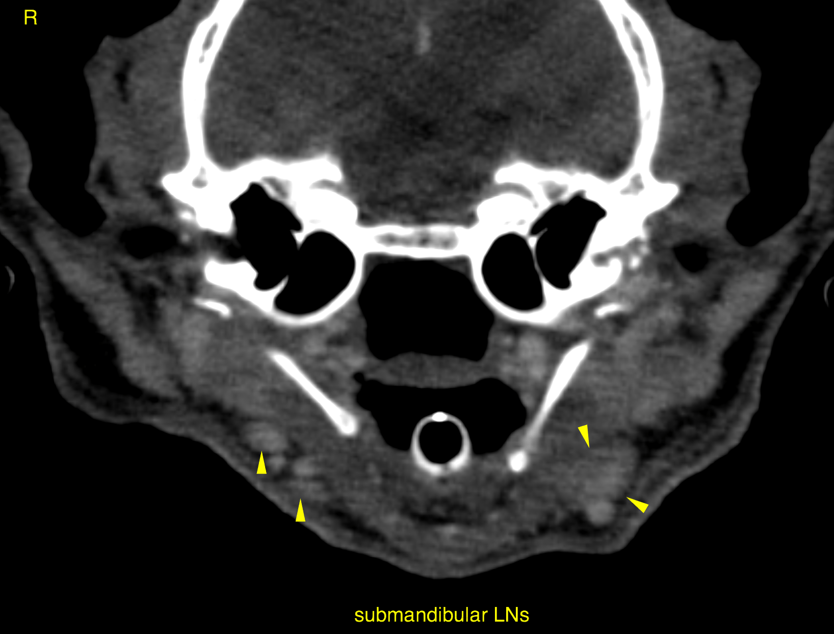
This 16 year old MN DSH cat presentd with possible squamous cell carcinoma of the larynx and surrounding tissue.
Physical exam: increased expiratory sounds
This 16 year old MN DSH cat presentd with possible squamous cell carcinoma of the larynx and surrounding tissue.
Physical exam: increased expiratory sounds