CT – Left-sided chronic diaphragmatic rupture in a 6 year old FS chihuahua mix

Case Study

CT – Left-sided chronic diaphragmatic rupture in a 6 year old FS chihuahua mix

This 6 year old FS Chihuahua mix dog presented for pre-surgical evaluation of a chronic diaphragmatic hernia

This 6 year old FS Chihuahua mix dog presented for pre-surgical evaluation of a chronic diaphragmatic hernia

DX

left sided diaphragmatic rupture

Image Interpretation

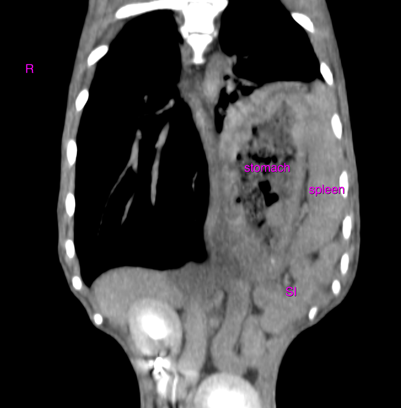
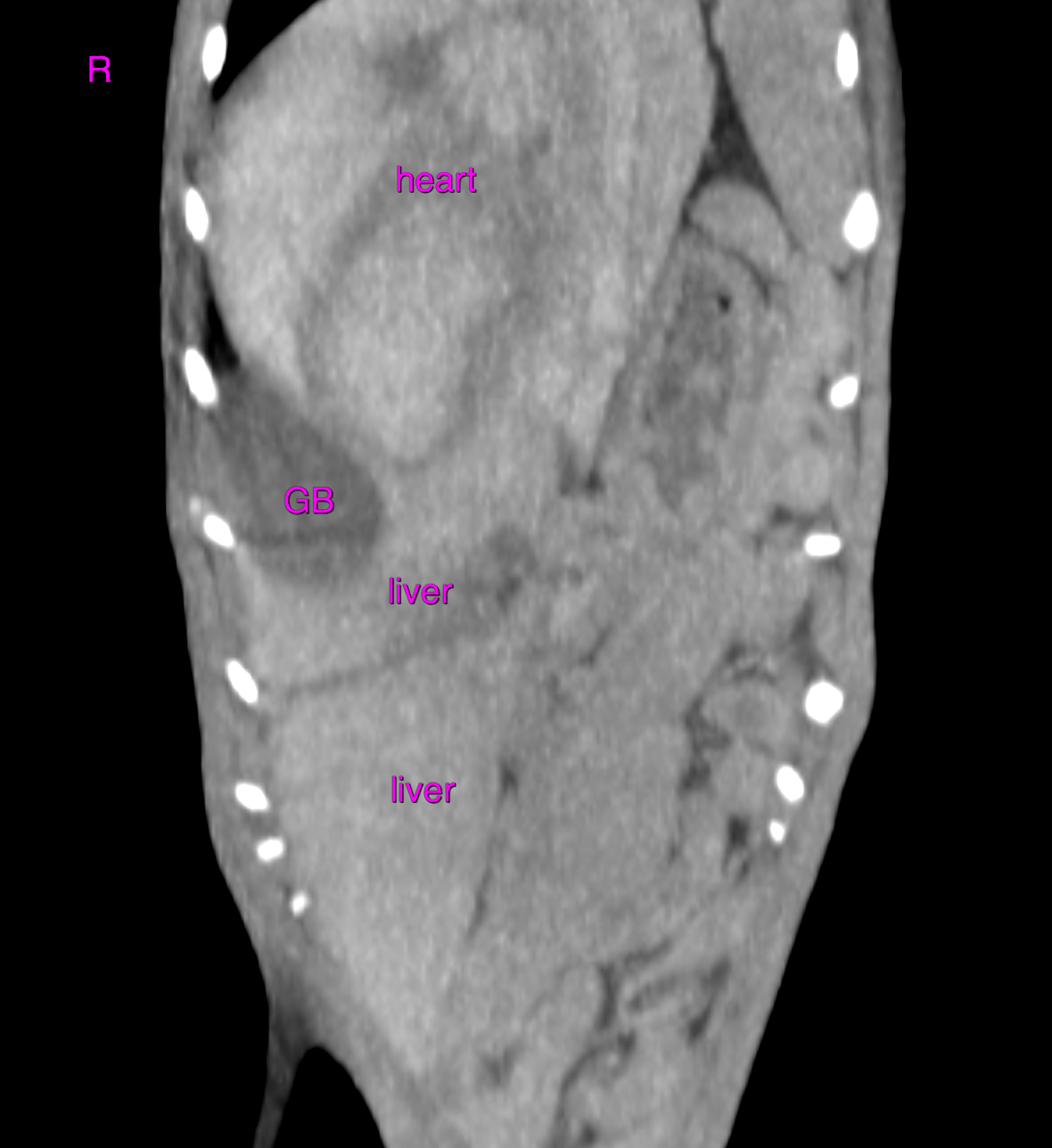
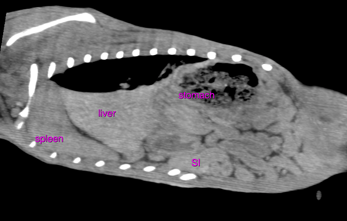
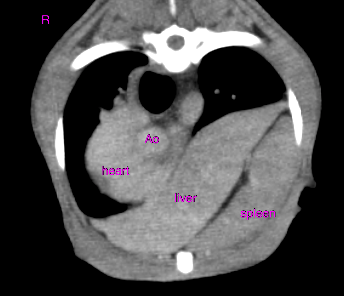
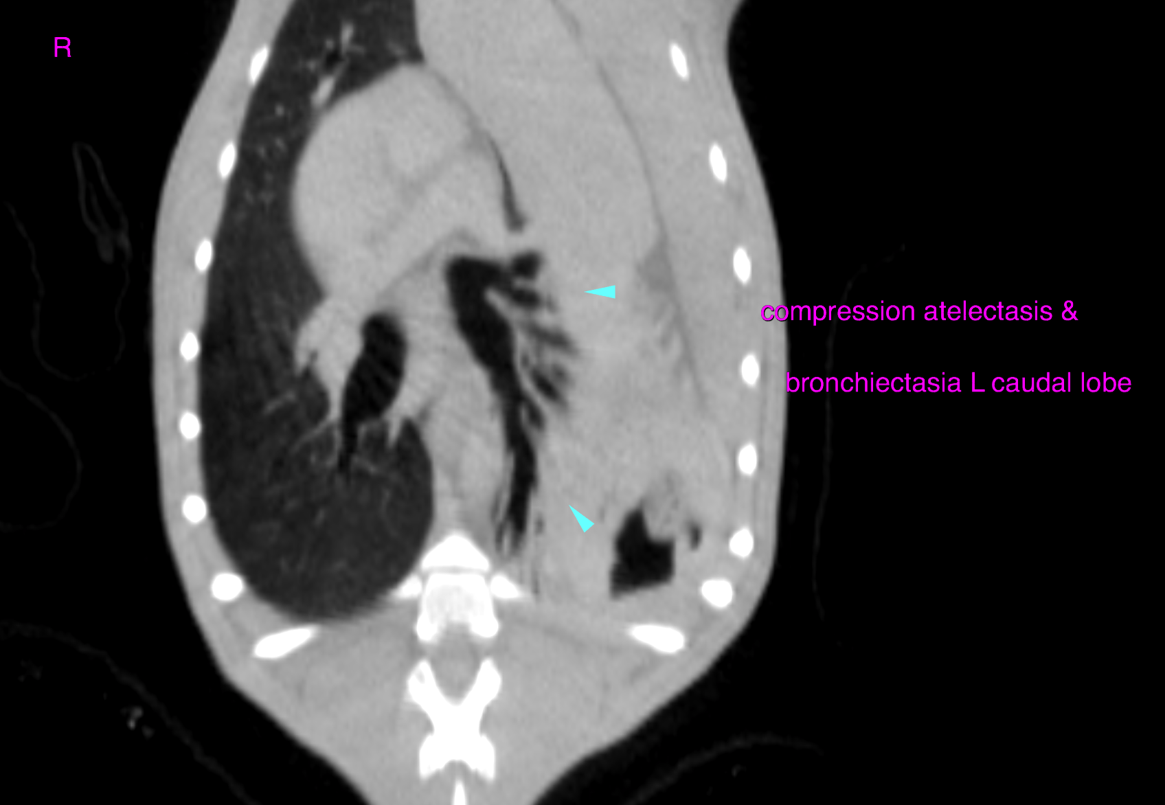
CT of the thorax and abdomen –  The spleen, stomach, omentum, part of the liver, pancreas, gallbladder and small intestine are herniated into the left hemithorax through a large mainly left-sided diaphragmatic defect. The mass effect of the herniated organs leads to a right-sided mediastinal shift with deviation of the heart and compression atelectasis of the left caudal lung lobe. Generalized mid bronchiectasia is noted and likely to be a function of the breathhold technique.  The organs of the urinary tract, part of the liver and intestine are left within the abdomen. The abdominal wall is tucked up.

Outcome

At the time of the examination no differential perfusion of the liver or splenic parenchyma was noted and there was no evidence of gastric dilation which are possible signs of organ incarceration. However, elective surgery should be considered as spontaneous organ incarceration is a possibly fatal complication of diaphragmatic rupture and is commonly associated with presentation as an emergency case. The anatomical reconstruction of the diaphragm may be complicated by cicatricial retraction of the ruptured diaphragm

Patient Information

Patient Name : Dixie Skoulikas/MPI
Gender : Female, Spayed
Species : Canine
Type of Imaging : Ultrasound
Status : Complete
Liz Wuz Here : Yes
Code : 11_00064

Images

bildschirmfoto_2016-06-29_um_13bildschirmfoto_2016-06-29_um_13bildschirmfoto_2016-06-29_um_13bildschirmfoto_2016-06-29_um_13bildschirmfoto_2016-06-29_um_13
Skip to content