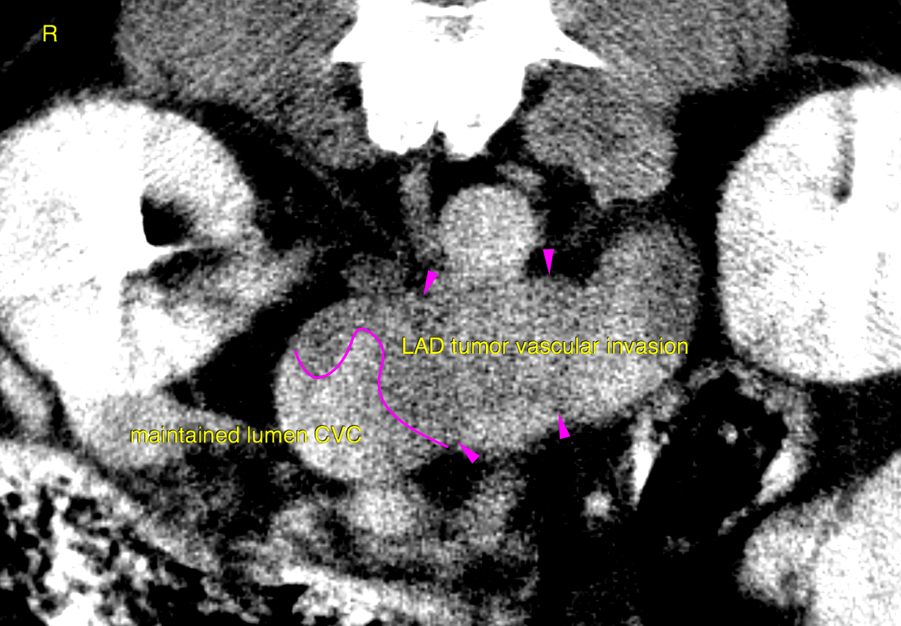
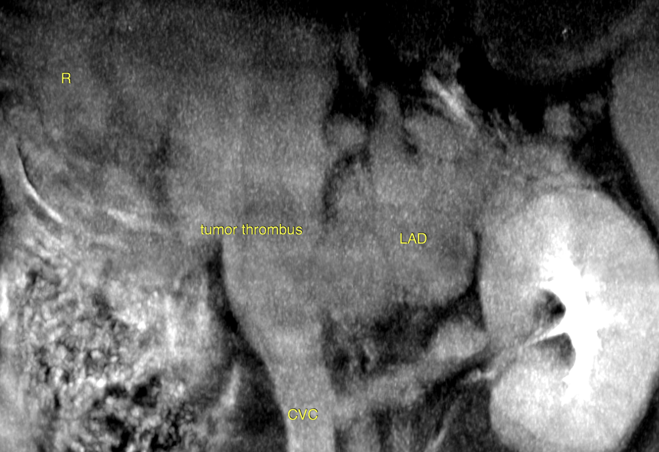
CT of the abdomen – A leftsided polygonal expansile and slightly ill defined adrenal mass of 4 x 3.5 x 4 cm is seen. The mass reveals extensive invasion into the caudal vena cava obstructing 60 % of the vascular diameter and expanding the vascular lumen signifcantly. The intravascular tumor extends at least 7 cm cranial from the right renal vein. Marginal blood flow is maintained. Pooling of contrast caudal to the vascular mass with differential contrast concentration is seen. The mass itself reveals a heterogenous attenuation pattern with non-uniform contrast enhancement. Mild localized retroperitoneal effusion and fat stranding is noted surrounding the mass.
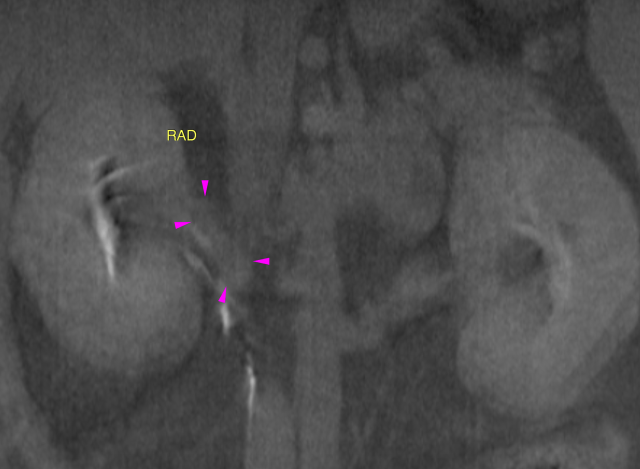
The right adrenal gland assessment is limited by artifacts. However the cranial and caudal pole diameter is less than 0.55 cm. The regular organ architecture is maintained.