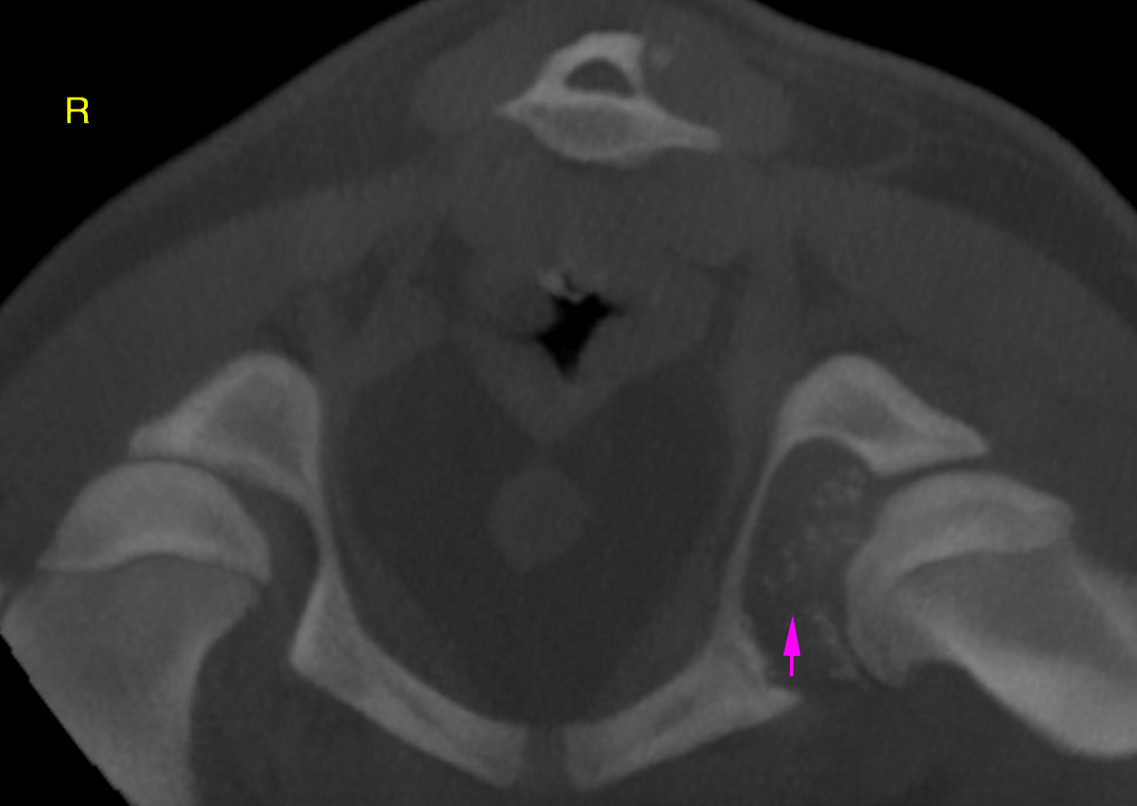
CT – Hereditary congenital multiple cartilaginous exostosis with compressive myelopathy and neuropathy in a 5 month old M Border Collie

Case Study

CT – Hereditary congenital multiple cartilaginous exostosis with compressive myelopathy and neuropathy in a 5 month old M Border Collie

This 5 month old M Border Collie dog presented with a Wobbler-like gait, difficulty rising. Muscle atrophy right hind but reflexes present, some TL discomfort. No neck mobility. Exostosis present in multiple areas. 

This 5 month old M Border Collie dog presented with a Wobbler-like gait, difficulty rising. Muscle atrophy right hind but reflexes present, some TL discomfort. No neck mobility. Exostosis present in multiple areas. 

DX

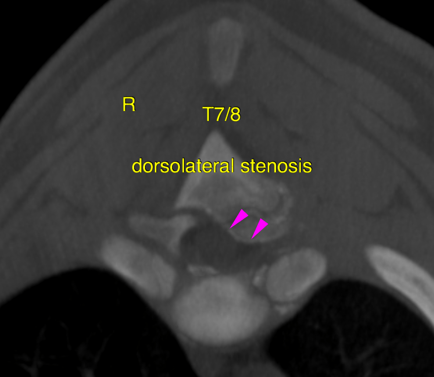
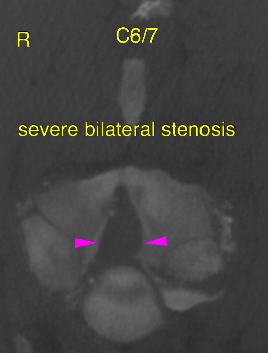
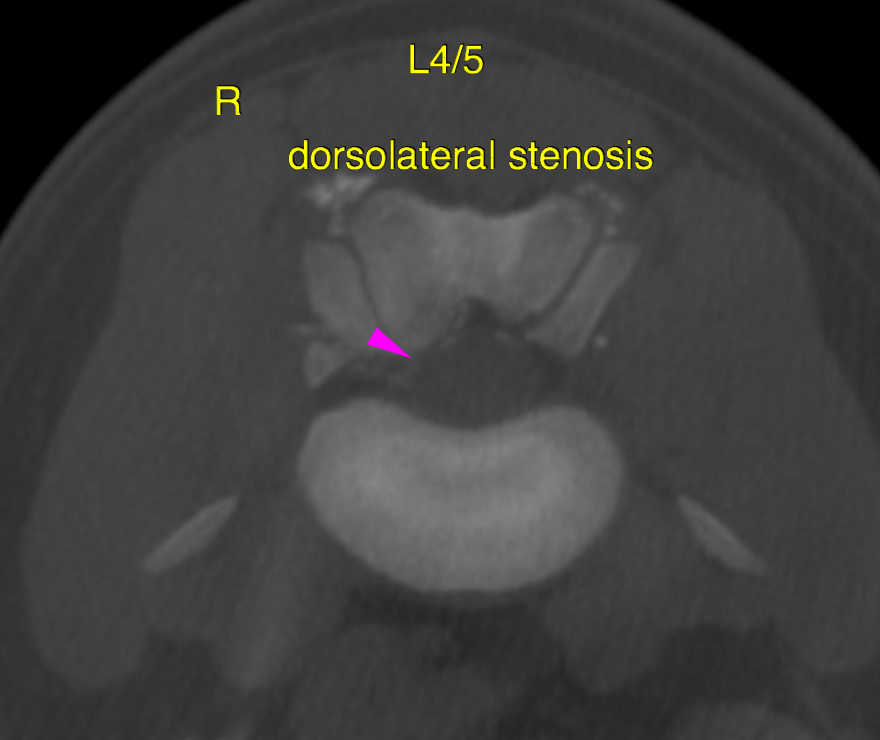
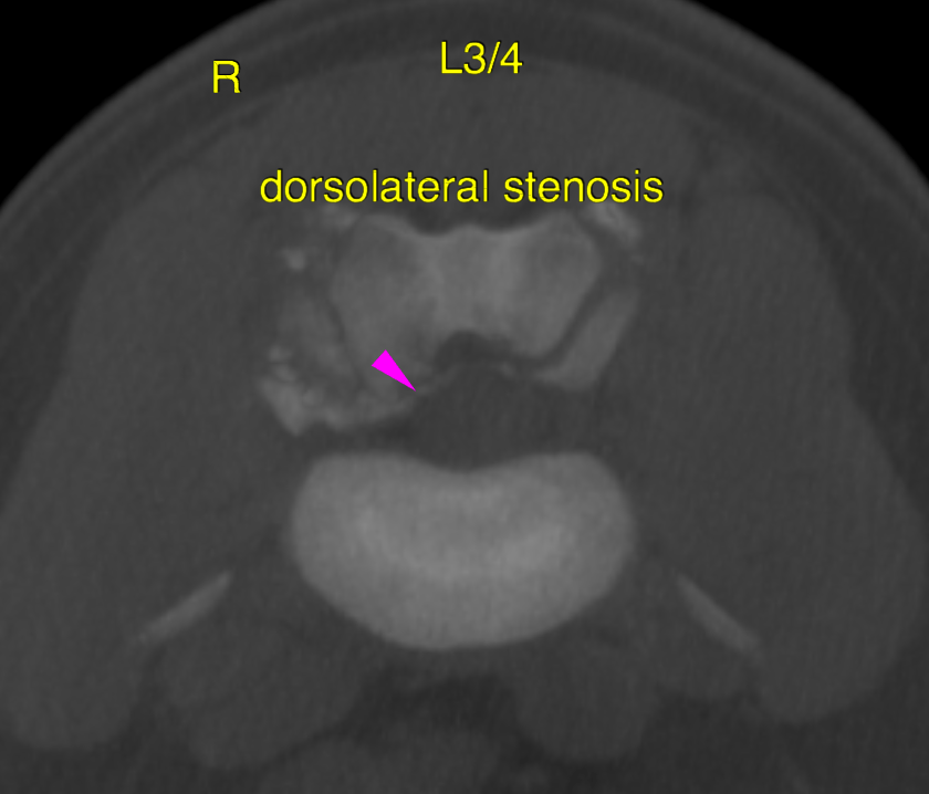
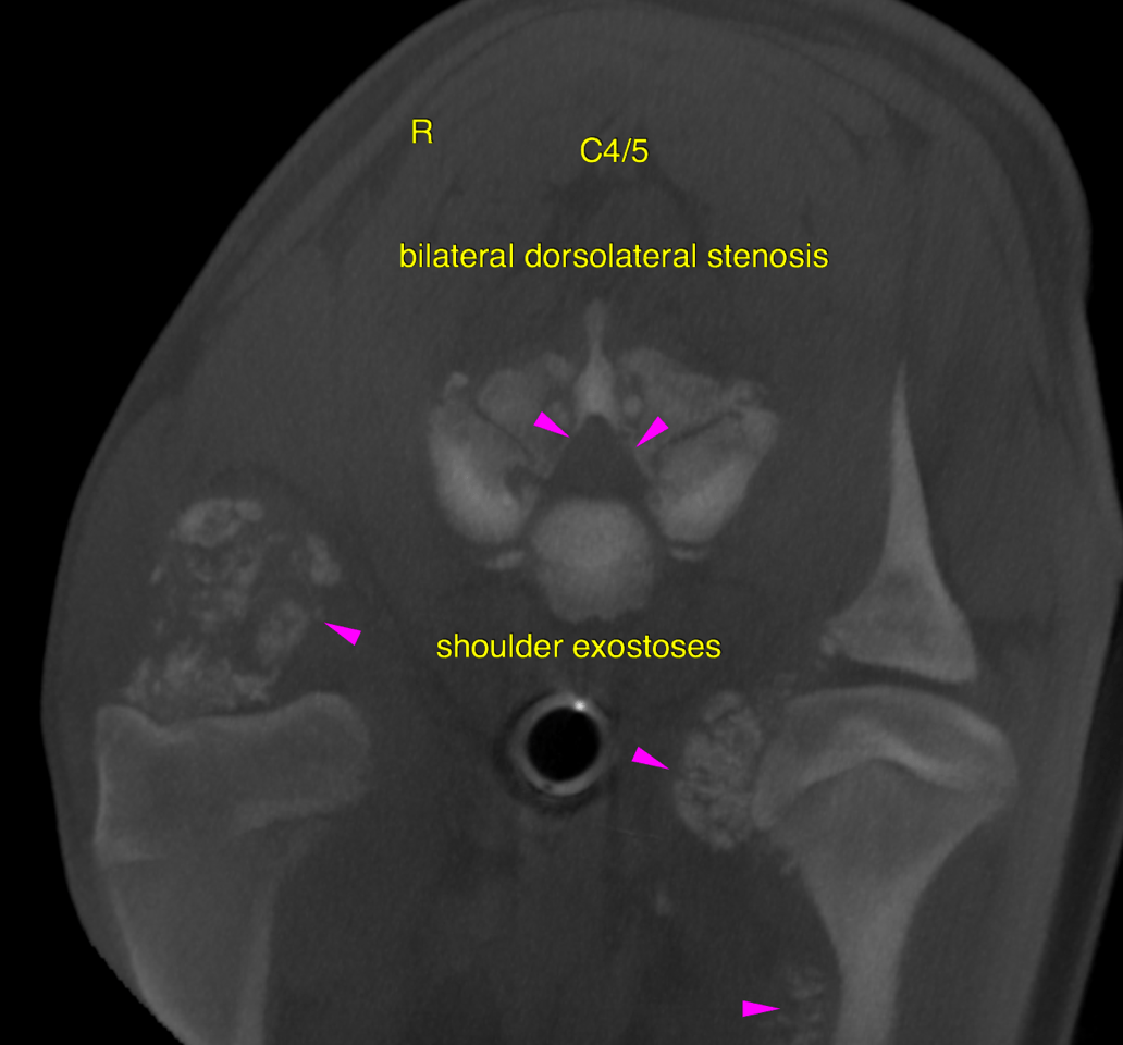
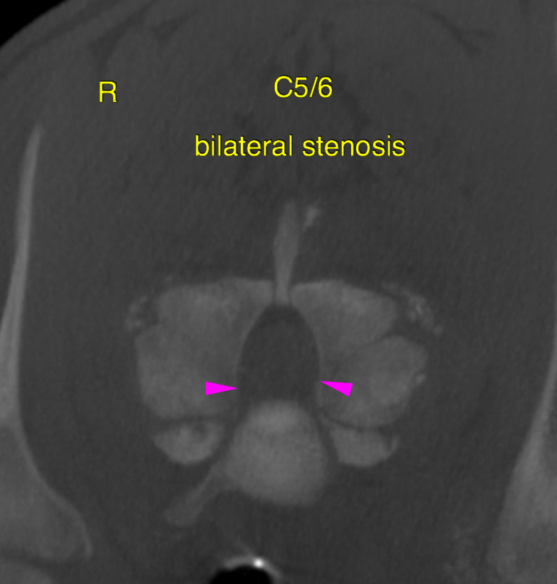
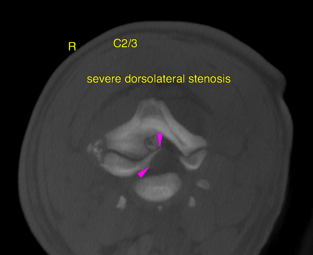
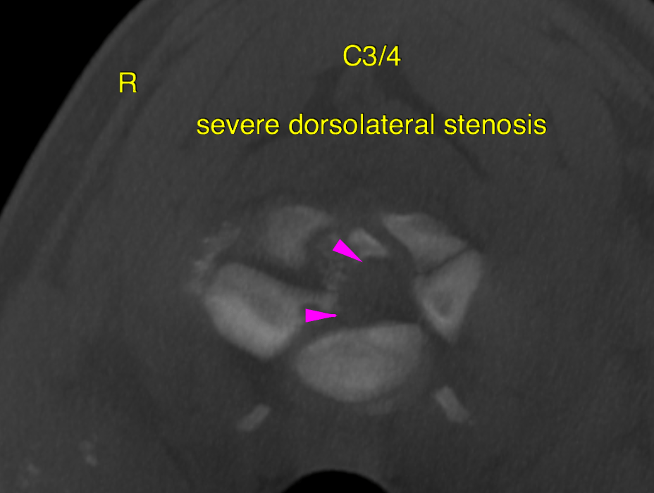
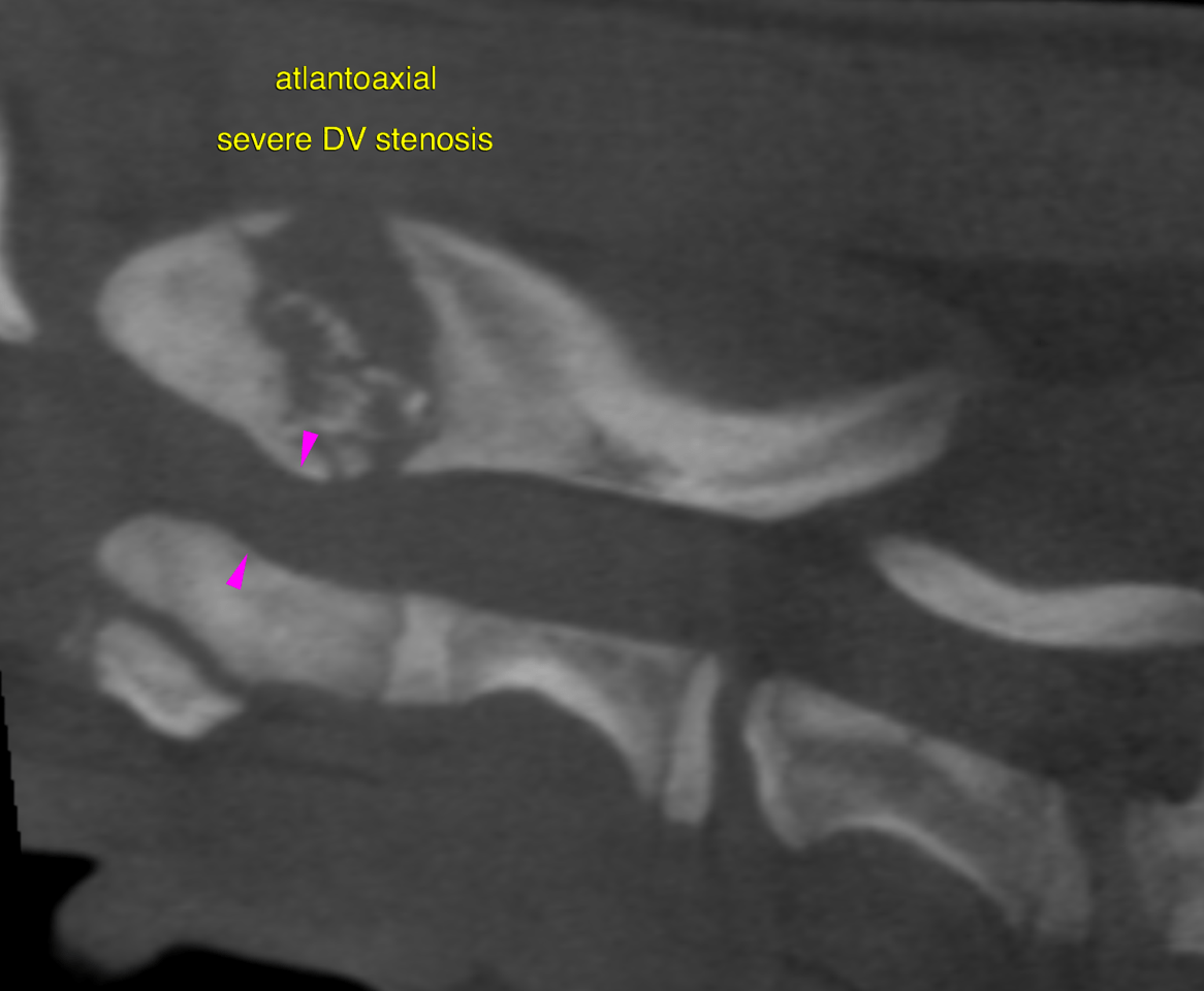
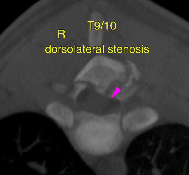
The computed tomographic findings of the spine are compatible with the history of hereditary congenital multiple cartilaginous exostosis. Multifocal moderate to severe vertebral canal and neuroforaminal stenosis is present and assumed to cause compressive myelopathy and neuropathy. Partial fusion of the intervertebral transition is evident as well and likely to be associated with restricted range of motion.

Image Interpretation

CT of the spine – The computed tomography of the spine reveals multiple sites of expansile stippled new bone formation covered by cartilage-attenuating tissue associated with the articular and epiphyseal bones. Multiple vertebral joint facets, vertebral arches, spinous processes, vertebral endplates, articulations of the ribs to the vertebrae are affected as well as the scapulae, humeri and left femoral head. The lesions bridge the neighboring vertebrae. They are space occupying and associated with multifocal facet joint hyperplasia resulting in multifocal moderate to severe vertebral canal stenosis and neuroforaminal exit zone stenosis. Marked reduction of the cross sectional area of the vertebral canal is noted at the atlantoaaxial level as well as throughout the entire cervical spine, T7/8, T9/10, L3/4 and L5/6. Multifocal compressive myelopathy and neuropathy has to be assumed. Faint focal mineralization of the intraabdominal vasculature is noted and likely to be a function of the primary disease.
Outcome

Malignant transformation of the osteomatous lesions is a potential, but rare.
In general surgical resection of the osteomas is a possible treatment option when
clinical signs occur. However, this option is very limited here owing to the multifocal
and serial stenosis throughout the spine – especially in the cervical area.

Patient Information

Patient Name : Benji Skoyec/CVC
Gender : Male, Intact
Species : Canine
Type of Imaging : Ultrasound
Status : Complete
Liz Wuz Here : Yes
Code : 16_00022

Exam Finding

  • Ambulatory deficits
  • Neurologic signs

Images

bildschirmfoto_2016-05-24_um_10bildschirmfoto_2016-05-24_um_10bildschirmfoto_2016-05-24_um_10bildschirmfoto_2016-05-24_um_10bildschirmfoto_2016-05-24_um_10bildschirmfoto_2016-05-24_um_10bildschirmfoto_2016-05-24_um_10bildschirmfoto_2016-05-24_um_10bildschirmfoto_2016-05-24_um_10bildschirmfoto_2016-05-24_um_10bildschirmfoto_2016-05-24_um_10
Skip to content